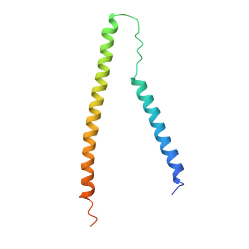
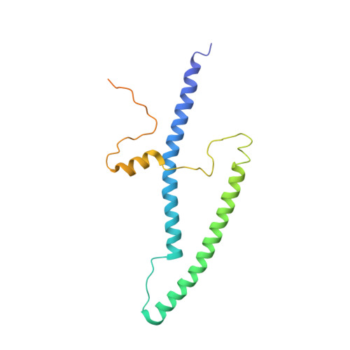
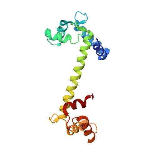
1YTZ

Crystal structure of skeletal muscle troponin in the Ca2+-activated state


Experimental Data Snapshot

  • Method: X-RAY DIFFRACTION
  • Resolution: 3.00 Å
  • R-Value Free: 0.338 
  • R-Value Work: 0.289 
  • R-Value Observed: 0.289 

wwPDB Validation   3D Report Full Report


Ligand Structure Quality Assessment 


This is version 1.4 of the entry. See complete history


Literature

Ca2+-regulated structural changes in troponin

Vinogradova, M.V.Stone, D.B.Malanina, G.G.Karatzaferi, C.Cooke, R.Mendelson, R.A.Fletterick, R.J.

(2005) Proc Natl Acad Sci U S A 102: 5038-5043

  • DOI: https://doi.org/10.1073/pnas.0408882102
  • Primary Citation of Related Structures:  
    1YTZ, 1YV0

  • PubMed Abstract: 

    Troponin senses Ca2+ to regulate contraction in striated muscle. Structures of skeletal muscle troponin composed of TnC (the sensor), TnI (the regulator), and TnT (the link to the muscle thin filament) have been determined. The structure of troponin in the Ca(2+)-activated state features a nearly twofold symmetrical assembly of TnI and TnT subunits penetrated asymmetrically by the dumbbell-shaped TnC subunit. Ca ions are thought to regulate contraction by controlling the presentation to and withdrawal of the TnI inhibitory segment from the thin filament. Here, we show that the rigid central helix of the sensor binds the inhibitory segment of TnI in the Ca(2+)-activated state. Comparison of crystal structures of troponin in the Ca(2+)-activated state at 3.0 angstroms resolution and in the Ca(2+)-free state at 7.0 angstroms resolution shows that the long framework helices of TnI and TnT, presumed to be a Ca(2+)-independent structural domain of troponin are unchanged. Loss of Ca ions causes the rigid central helix of the sensor to collapse and to release the inhibitory segment of TnI. The inhibitory segment of TnI changes conformation from an extended loop in the presence of Ca2+ to a short alpha-helix in its absence. We also show that Anapoe, a detergent molecule, increases the contractile force of muscle fibers and binds specifically, together with the TnI switch helix, in a hydrophobic pocket of TnC upon activation by Ca ions.


  • Organizational Affiliation

    Department of Biochemistry and Biophysics, University of California, San Francisco, CA 94143-2240, USA.


Macromolecules
Find similar proteins by:  (by identity cutoff)  |  3D Structure
Entity ID: 1
MoleculeChains Sequence LengthOrganismDetailsImage
Troponin TA [auth T]107Gallus gallusMutation(s): 0 
UniProt
Find proteins for P12620 (Gallus gallus)
Explore P12620 
Go to UniProtKB:  P12620
Entity Groups  
Sequence Clusters30% Identity50% Identity70% Identity90% Identity95% Identity100% Identity
UniProt GroupP12620
Sequence Annotations
Expand
  • Reference Sequence
Find similar proteins by:  (by identity cutoff)  |  3D Structure
Entity ID: 2
MoleculeChains Sequence LengthOrganismDetailsImage
Troponin IB [auth I]182Gallus gallusMutation(s): 2 
UniProt
Find proteins for P68246 (Gallus gallus)
Explore P68246 
Go to UniProtKB:  P68246
Entity Groups  
Sequence Clusters30% Identity50% Identity70% Identity90% Identity95% Identity100% Identity
UniProt GroupP68246
Sequence Annotations
Expand
  • Reference Sequence
Find similar proteins by:  (by identity cutoff)  |  3D Structure
Entity ID: 3
MoleculeChains Sequence LengthOrganismDetailsImage
Troponin C162Gallus gallusMutation(s): 0 
Gene Names: TNNC2
UniProt
Find proteins for P02588 (Gallus gallus)
Explore P02588 
Go to UniProtKB:  P02588
Entity Groups  
Sequence Clusters30% Identity50% Identity70% Identity90% Identity95% Identity100% Identity
UniProt GroupP02588
Sequence Annotations
Expand
  • Reference Sequence
Experimental Data & Validation

Experimental Data

  • Method: X-RAY DIFFRACTION
  • Resolution: 3.00 Å
  • R-Value Free: 0.338 
  • R-Value Work: 0.289 
  • R-Value Observed: 0.289 
  • Space Group: P 43 21 2
Unit Cell:
Length ( Å )Angle ( ˚ )
a = 138.608α = 90
b = 138.608β = 90
c = 83.676γ = 90
Software Package:
Software NamePurpose
DENZOdata reduction
SCALEPACKdata scaling
CNSrefinement
CNSphasing

Structure Validation

View Full Validation Report


Ligand Structure Quality Assessment 


Entry History 

Deposition Data

Revision History  (Full details and data files)

  • Version 1.0: 2005-04-12
    Type: Initial release
  • Version 1.1: 2008-04-30
    Changes: Version format compliance
  • Version 1.2: 2011-07-13
    Changes: Version format compliance
  • Version 1.3: 2021-10-20
    Changes: Database references, Derived calculations, Structure summary
  • Version 1.4: 2024-02-14
    Changes: Data collection