Redox-dependent structural differences in putidaredoxin derived from homologous structure refinement via residual dipolar couplings.
Jain, N.U., Tjioe, E., Savidor, A., Boulie, J.(2005) Biochemistry 44: 9067-9078
- PubMed: 15966730 Search on PubMed
- DOI: https://doi.org/10.1021/bi050152c
- Primary Citation Related Structures:
1YJI, 1YJJ - PubMed Abstract:
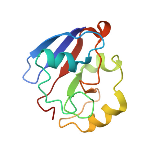
Structural differences in the [2Fe-2S] ferredoxin, putidaredoxin (Pdx), from the camphor hydroxylation pathway of Pseudomonas putida have been investigated as a function of oxidation state of the iron cluster. Pdx is involved in biological electron transfer to cytochrome P450(cam) (CYP101). Redox-dependent differences have been observed previously for Pdx in terms of binding affinities to CYP101, NMR spectral differences, and dynamic properties. To further characterize these differences, structure refinement of both oxidized and reduced Pdx has been carried out using a hybrid approach utilizing paramagnetic distance restraints and NMR orientational restraints in the form of backbone (15)N residual dipolar couplings. Use of these new restraints has improved the structure of oxidized Pdx considerably over the earlier solution NMR structure without RDC restraints, with the new structure now much closer in overall fold to the recently published X-ray crystal structures. We now observe better defined relative orientations of the major secondary structure elements as also of the conformation of the metal binding loop region. Extension of this approach to structure calculation of reduced Pdx has identified structural differences that are primarily localized for residues in the C-terminal interaction domain consisting of the functionally important residue Trp 106 and regions near the metal binding loop in Pdx. These redox-dependent structural differences in Pdx correlate to dynamic changes observed before and may be linked to differences in binding and electron transfer properties between oxidized and reduced Pdx.
- Biochemistry, Cellular and Molecular Biology Department, University of Tennessee, Knoxville, Tennessee 37996-0840, USA. njain@utk.edu
Organizational Affiliation: