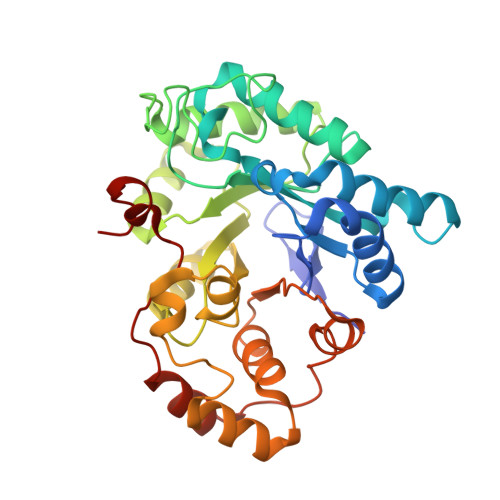
1YE4

Crystal structure of the Lys-274 to Arg mutant of Candida tenuis xylose reductase (AKR2B5) bound to NAD+


Experimental Data Snapshot

  • Method: X-RAY DIFFRACTION
  • Resolution: 2.40 Å
  • R-Value Free: 0.256 
  • R-Value Work: 0.206 
  • R-Value Observed: 0.206 

Starting Model: experimental
View more details

wwPDB Validation   3D Report Full Report


Ligand Structure Quality Assessment 


This is version 1.4 of the entry. See complete history


Literature

Fine tuning of coenzyme specificity in family 2 aldo-keto reductases revealed by crystal structures of the Lys-274-->Arg mutant of Candida tenuis xylose reductase (AKR2B5) bound to NAD(+) and NADP(+).

Leitgeb, S.Petschacher, B.Wilson, D.K.Nidetzky, B.

(2005) FEBS Lett 579: 763-767

  • DOI: https://doi.org/10.1016/j.febslet.2004.12.063
  • Primary Citation of Related Structures:  
    1YE4, 1YE6

  • PubMed Abstract: 

    Aldo-keto reductases of family 2 employ single site replacement Lys-->Arg to switch their cosubstrate preference from NADPH to NADH. X-ray crystal structures of Lys-274-->Arg mutant of Candida tenuis xylose reductase (AKR2B5) bound to NAD+ and NADP+ were determined at a resolution of 2.4 and 2.3A, respectively. Due to steric conflicts in the NADP+-bound form, the arginine side chain must rotate away from the position of the original lysine side chain, thereby disrupting a network of direct and water-mediated interactions between Glu-227, Lys-274 and the cofactor 2'-phosphate and 3'-hydroxy groups. Because anchoring contacts of its Glu-227 are lost, the coenzyme-enfolding loop that becomes ordered upon binding of NAD(P)+ in the wild-type remains partly disordered in the NADP+-bound mutant. The results delineate a catalytic reaction profile for the mutant in comparison to wild-type.


  • Organizational Affiliation

    Institute of Biotechnology and Biochemical Engineering, Graz University of Technology, Petersgasse 12/I, A-8010 Graz, Austria.


Macromolecules
Find similar proteins by:  (by identity cutoff)  |  3D Structure
Entity ID: 1
MoleculeChains Sequence LengthOrganismDetailsImage
NAD(P)H-dependent D-xylose reductase
A, B, C, D
322Yamadazyma tenuisMutation(s): 1 
Gene Names: XYL1XYLR
EC: 1.1.1
UniProt
Find proteins for O74237 (Candida tenuis)
Explore O74237 
Go to UniProtKB:  O74237
Entity Groups  
Sequence Clusters30% Identity50% Identity70% Identity90% Identity95% Identity100% Identity
UniProt GroupO74237
Sequence Annotations
Expand
  • Reference Sequence
Small Molecules
Ligands 2 Unique
IDChains Name / Formula / InChI Key2D Diagram3D Interactions
NAD
Query on NAD

Download Ideal Coordinates CCD File 
F [auth A],
I [auth B],
K [auth C],
N [auth D]
NICOTINAMIDE-ADENINE-DINUCLEOTIDE
C21 H27 N7 O14 P2
BAWFJGJZGIEFAR-NNYOXOHSSA-N
SO4
Query on SO4

Download Ideal Coordinates CCD File 
E [auth A]
G [auth B]
H [auth B]
J [auth C]
L [auth D]
E [auth A],
G [auth B],
H [auth B],
J [auth C],
L [auth D],
M [auth D]
SULFATE ION
O4 S
QAOWNCQODCNURD-UHFFFAOYSA-L
Experimental Data & Validation

Experimental Data

  • Method: X-RAY DIFFRACTION
  • Resolution: 2.40 Å
  • R-Value Free: 0.256 
  • R-Value Work: 0.206 
  • R-Value Observed: 0.206 
  • Space Group: C 1 2 1
Unit Cell:
Length ( Å )Angle ( ˚ )
a = 182.827α = 90
b = 128.666β = 90.11
c = 80.101γ = 90
Software Package:
Software NamePurpose
SCALEPACKdata scaling
CNSrefinement
DENZOdata reduction
CNSphasing

Structure Validation

View Full Validation Report


Ligand Structure Quality Assessment 


Entry History 

Deposition Data

Revision History  (Full details and data files)

  • Version 1.0: 2005-02-01
    Type: Initial release
  • Version 1.1: 2008-04-30
    Changes: Version format compliance
  • Version 1.2: 2011-07-13
    Changes: Version format compliance
  • Version 1.3: 2021-10-20
    Changes: Database references, Derived calculations
  • Version 1.4: 2023-08-23
    Changes: Data collection, Refinement description