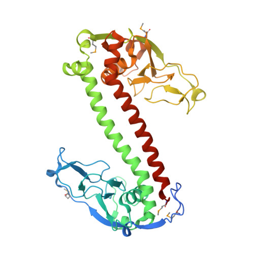
Crystal Structure of a Putative Type I Restriction-Modification S Subunit from Mycoplasma genitalium
Calisto, B.M., Pich, O.Q., Pinol, J., Fita, I., Querol, E., Carpena, X.(2005) J Mol Biology 351: 749-762
- PubMed: 16038930
- DOI: https://doi.org/10.1016/j.jmb.2005.06.050
- Primary Citation of Related Structures:
1YDX - PubMed Abstract:
The crystal structure of the eubacteria Mycoplasma genitalium ORF MG438 polypeptide, determined by multiple anomalous dispersion and refined at 2.3 A resolution, reveals the organization of S subunits from the Type I restriction and modification system. The structure consists of two globular domains, with about 150 residues each, separated by a pair of 40 residue long antiparallel alpha-helices. The globular domains correspond to the variable target recognition domains (TRDs), as previously defined for S subunits on sequence analysis, while the two helices correspond to the central (CR1) and C-terminal (CR2) conserved regions, respectively. The structure of the MG438 subunit presents an overall cyclic topology with an intramolecular 2-fold axis that superimposes the N and the C-half parts, each half containing a globular domain and a conserved helix. TRDs are found to be structurally related with the small domain of the Type II N6-adenine DNA MTase TaqI. These relationships together with the structural peculiarities of MG438, in particular the presence of the intramolecular quasi-symmetry, allow the proposal of a model for S subunits recognition of their DNA targets in agreement with previous experimental results. In the crystal, two subunits of MG438 related by a crystallographic 2-fold axis present a large contact area mainly involving the symmetric interactions of a cluster of exposed hydrophobic residues. Comparison with the recently reported structure of an S subunit from the archaea Methanococcus jannaschii highlights the structural features preserved despite a sequence identity below 20%, but also reveals important differences in the globular domains and in their disposition with respect to the conserved regions.
- Institut de Biologia Molecular de Barcelona (IBMB-CSIC), Parc Científic de Barcelona, Josep-Samitier 1-5, 08028 Barcelona, Spain.
Organizational Affiliation:


