Structure and lytic activity of a Bacillus anthracis prophage endolysin
Low, L.Y., Yang, C., Perego, M., Osterman, A., Liddington, R.C.(2005) J Biological Chem 280: 35433-35439
- PubMed: 16103125 Search on PubMed
- DOI: https://doi.org/10.1074/jbc.M502723200
- Primary Citation Related Structures:
1YB0, 2AR3 - PubMed Abstract:
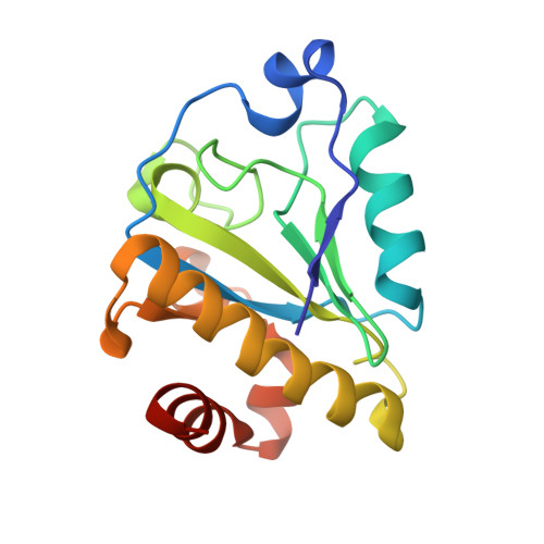
We report a structural and functional analysis of the lambda prophage Ba02 endolysin (PlyL) encoded by the Bacillus anthracis genome. We show that PlyL comprises two autonomously folded domains, an N-terminal catalytic domain and a C-terminal cell wall-binding domain. We determined the crystal structure of the catalytic domain; its three-dimensional fold is related to that of the cell wall amidase, T7 lysozyme, and contains a conserved zinc coordination site and other components of the catalytic machinery. We demonstrate that PlyL is an N-acetylmuramoyl-L-alanine amidase that cleaves the cell wall of several Bacillus species when applied exogenously. We show, unexpectedly, that the catalytic domain of PlyL cleaves more efficiently than the full-length protein, except in the case of Bacillus cereus, and using GFP-tagged cell wall-binding domain, we detected strong binding of the cell wall-binding domain to B. cereus but not to other species tested. We further show that a related endolysin (Ply21) from the B. cereus phage, TP21, shows a similar pattern of behavior. To explain these data, and the species specificity of PlyL, we propose that the C-terminal domain inhibits the activity of the catalytic domain through intramolecular interactions that are relieved upon binding of the C-terminal domain to the cell wall. Furthermore, our data show that (when applied exogenously) targeting of the enzyme to the cell wall is not a prerequisite of its lytic activity, which is inherently high. These results may have broad implications for the design of endolysins as therapeutic agents.
- Infectious and Inflammatory Disease Center, The Burnham Institute, La Jolla, California 92037, USA.
Organizational Affiliation: