1YAR

Structure of Archeabacterial 20S proteasome mutant D9S- PA26 complex


Experimental Data Snapshot

  • Method: X-RAY DIFFRACTION
  • Resolution: 1.90 Å
  • R-Value Free: 0.216 
  • R-Value Work: 0.182 
  • R-Value Observed: 0.182 

wwPDB Validation   3D Report Full Report


This is version 1.4 of the entry. See complete history


Literature

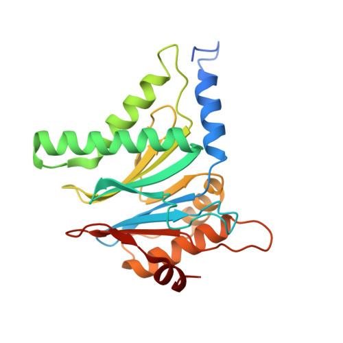
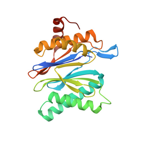
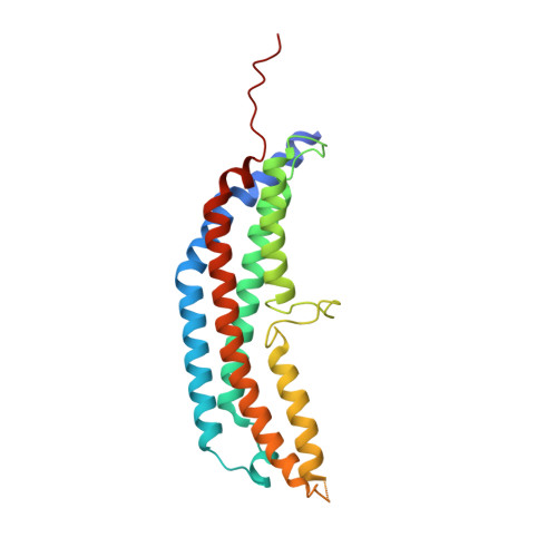
The 1.9 A structure of a proteasome-11S activator complex and implications for proteasome-PAN/PA700 interactions.

Forster, A.Masters, E.I.Whitby, F.G.Robinson, H.Hill, C.P.

(2005) Mol Cell 18: 589-599

  • DOI: https://doi.org/10.1016/j.molcel.2005.04.016
  • Primary Citation of Related Structures:  
    1YA7, 1YAR, 1YAU, 1Z7Q

  • PubMed Abstract: 

    Proteasomes are cylindrical structures that function in multiple cellular processes by degrading a wide variety of cytosolic and nuclear proteins. Substrate access and product release from the enclosed catalytic chamber occurs through axial pores that are opened by activator complexes. Here, we report high-resolution structures of wild-type and mutant archaeal proteasomes bound to the activator PA26. These structures support the proposal that an ordered open conformation is required for proteolysis and that its formation can be triggered by outward displacement of surrounding residues. The structures and associated biochemical assays reveal the mechanism of binding, which involves an interaction between the PA26 C terminus and a conserved lysine. Surprisingly, biochemical observations implicate an equivalent interaction for the unrelated ATP-dependent activators PAN and PA700.


  • Organizational Affiliation

    Department of Biochemistry, University of Utah School of Medicine, Salt Lake City, Utah 84132, USA.


Macromolecules
Find similar proteins by:  (by identity cutoff)  |  3D Structure
Entity ID: 1
MoleculeChains Sequence LengthOrganismDetailsImage
Proteasome alpha subunit
A, B, C, D, E
A, B, C, D, E, F, G
233Thermoplasma acidophilumMutation(s): 1 
Gene Names: psmA
EC: 3.4.25.1
UniProt
Find proteins for P25156 (Thermoplasma acidophilum (strain ATCC 25905 / DSM 1728 / JCM 9062 / NBRC 15155 / AMRC-C165))
Explore P25156 
Go to UniProtKB:  P25156
Entity Groups  
Sequence Clusters30% Identity50% Identity70% Identity90% Identity95% Identity100% Identity
UniProt GroupP25156
Sequence Annotations
Expand
  • Reference Sequence
Find similar proteins by:  (by identity cutoff)  |  3D Structure
Entity ID: 2
MoleculeChains Sequence LengthOrganismDetailsImage
Proteasome beta subunit
H, I, J, K, L
H, I, J, K, L, M, N
217Thermoplasma acidophilumMutation(s): 0 
Gene Names: psmB
EC: 3.4.25.1
UniProt
Find proteins for P28061 (Thermoplasma acidophilum (strain ATCC 25905 / DSM 1728 / JCM 9062 / NBRC 15155 / AMRC-C165))
Explore P28061 
Go to UniProtKB:  P28061
Entity Groups  
Sequence Clusters30% Identity50% Identity70% Identity90% Identity95% Identity100% Identity
UniProt GroupP28061
Sequence Annotations
Expand
  • Reference Sequence
Find similar proteins by:  (by identity cutoff)  |  3D Structure
Entity ID: 3
MoleculeChains Sequence LengthOrganismDetailsImage
proteasome activator protein PA26
O, P, Q, R, S
O, P, Q, R, S, T, U
237Trypanosoma bruceiMutation(s): 1 
UniProt
Find proteins for Q9U8G2 (Trypanosoma brucei)
Explore Q9U8G2 
Go to UniProtKB:  Q9U8G2
Entity Groups  
Sequence Clusters30% Identity50% Identity70% Identity90% Identity95% Identity100% Identity
UniProt GroupQ9U8G2
Sequence Annotations
Expand
  • Reference Sequence
Small Molecules
Ligands 2 Unique
IDChains Name / Formula / InChI Key2D Diagram3D Interactions
SO4
Query on SO4

Download Ideal Coordinates CCD File 
AA [auth F]
BA [auth F]
CA [auth G]
DA [auth H]
EA [auth H]
AA [auth F],
BA [auth F],
CA [auth G],
DA [auth H],
EA [auth H],
GA [auth I],
IA [auth J],
JA [auth J],
LA [auth K],
MA [auth K],
OA [auth L],
PA [auth L],
RA [auth M],
SA [auth M],
UA [auth N],
V [auth A],
VA [auth N],
W [auth B],
X [auth B],
Y [auth C],
Z [auth D]
SULFATE ION
O4 S
QAOWNCQODCNURD-UHFFFAOYSA-L
GOL
Query on GOL

Download Ideal Coordinates CCD File 
FA [auth H]
HA [auth I]
KA [auth J]
NA [auth K]
QA [auth L]
FA [auth H],
HA [auth I],
KA [auth J],
NA [auth K],
QA [auth L],
TA [auth M],
WA [auth N]
GLYCEROL
C3 H8 O3
PEDCQBHIVMGVHV-UHFFFAOYSA-N
Experimental Data & Validation

Experimental Data

  • Method: X-RAY DIFFRACTION
  • Resolution: 1.90 Å
  • R-Value Free: 0.216 
  • R-Value Work: 0.182 
  • R-Value Observed: 0.182 
  • Space Group: C 1 2 1
Unit Cell:
Length ( Å )Angle ( ˚ )
a = 254.874α = 90
b = 127.512β = 92.45
c = 181.182γ = 90
Software Package:
Software NamePurpose
REFMACrefinement
DENZOdata reduction
SCALEPACKdata scaling
AMoREphasing

Structure Validation

View Full Validation Report


Entry History 

Deposition Data

Revision History  (Full details and data files)

  • Version 1.0: 2005-07-26
    Type: Initial release
  • Version 1.1: 2008-04-30
    Changes: Version format compliance
  • Version 1.2: 2011-07-13
    Changes: Derived calculations, Version format compliance
  • Version 1.3: 2021-10-20
    Changes: Database references, Derived calculations
  • Version 1.4: 2023-08-23
    Changes: Data collection, Refinement description