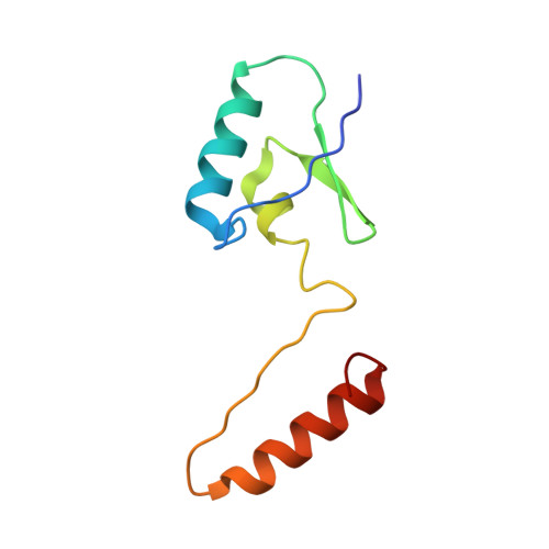
The HPr proteins from the thermophile Bacillus stearothermophilus can form domain-swapped dimers.
Sridharan, S., Razvi, A., Scholtz, J.M., Sacchettini, J.C.(2005) J Mol Biol 346: 919-931
- PubMed: 15713472 Search on PubMed
- DOI: https://doi.org/10.1016/j.jmb.2004.12.008
- Primary Citation Related Structures:
1Y4Y, 1Y50, 1Y51 - PubMed Abstract:
The study of proteins from extremophilic organisms continues to generate interest in the field of protein folding because paradigms explaining the enhanced stability of these proteins still elude us and such studies have the potential to further our knowledge of the forces stabilizing proteins. We have undertaken such a study with our model protein HPr from a mesophile, Bacillus subtilis, and a thermophile, Bacillus stearothermophilus. We report here the high-resolution structures of the wild-type HPr protein from the thermophile and a variant, F29W. The variant proved to crystallize in two forms: a monomeric form with a structure very similar to the wild-type protein as well as a domain-swapped dimer. Interestingly, the structure of the domain-swapped dimer for HPr is very different from that observed for a homologous protein, Crh, from B.subtilis. The existence of a domain-swapped dimer has implications for amyloid formation and is consistent with recent results showing that the HPr proteins can form amyloid fibrils. We also characterized the conformational stability of the thermophilic HPr proteins using thermal and solvent denaturation methods and have used the high-resolution structures in an attempt to explain the differences in stability between the different HPr proteins. Finally, we present a detailed analysis of the solution properties of the HPr proteins using a variety of biochemical and biophysical methods.
- Department of Biochemistry and Biophysics, Texas A&M University, College Station, TX 77843-2128, USA.
Organizational Affiliation: