1Y4C | pdb_00001y4c

Designed Helical Protein fusion MBP


Experimental Data Snapshot

  • Method: X-RAY DIFFRACTION
  • Resolution: 1.90 Å
  • R-Value Free: 
    0.225 (Depositor), 0.229 (DCC) 
  • R-Value Work: 
    0.202 (Depositor), 0.210 (DCC) 

wwPDB Validation 3D Report Full Report

Validation slider image for 1Y4C

This is version 2.1 of the entry. See complete history

Literature

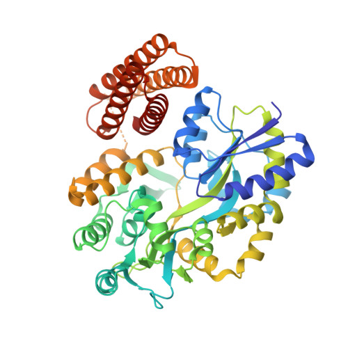
De novo design of an IL-4 antagonist and its structure at 1.9 A.

LaPorte, S.L.Forsyth, C.M.Cunningham, B.C.Miercke, L.J.Akhavan, D.Stroud, R.M.

(2005) Proc Natl Acad Sci U S A 102: 1889-1894

  • DOI: https://doi.org/10.1073/pnas.0408890102
  • Primary Citation Related Structures: 
    1Y4C

  • PubMed Abstract: 

    An IL-4 antagonist was designed based on structural and biochemical analysis of unbound IL-4 and IL-4 in complex with its high-affinity receptor (IL-4Ralpha). Our design strategy sought to capture a protein-protein interaction targeting the high affinity that IL-4 has for IL-4Ralpha. This strategy has impact due to the potential relevance of IL-4Ralpha as a drug target in the treatment of asthma. To mimic the IL-4 binding surface, critical side chains for receptor binding were identified, and these side chains were transplanted onto a previously characterized, de novo-designed four-helix protein called designed helical protein 1 (DHP-1). This first-generation design resolved the ambiguity previously described for the connectivity between helices in DHP-1 and resulted in a protein capable of binding to IL-4Ralpha. The second-generation antagonist was based upon further molecular modeling, and it succeeded in binding IL-4Ralpha better than the first-generation. This protein, termed DHP-14-AB, yielded a protein with a cooperative unfolding transition (DeltaGu0=8.1 kcal/mol) and an IC50 of 27 microM when in competition with IL-4 whereas DHP-1 had no affinity for IL-4Ralpha. The crystal structure of DHP-14-AB was determined to 1.9-A resolution and was compared with IL-4. This comparison revealed how design strategies targeting protein-protein interactions require high-resolution 3D data and the incorporation of orientation-specific information at the level of side-chains and secondary structure element interactions.


  • Organizational Affiliation
    • Department of Biochemistry and Biophysics, University of California, 600 16th Street, Box 2240, San Francisco, CA 94143-2240, USA.

Macromolecule Content 

  • Total Structure Weight: 54.63 kDa 
  • Atom Count: 3,979 
  • Modeled Residue Count: 480 
  • Deposited Residue Count: 494 
  • Unique protein chains: 1

Macromolecules

Find similar proteins by:|  3D Structure
Entity ID: 1
MoleculeChains  Sequence LengthOrganismDetailsImage
Maltose binding protein fused with designed helical protein494Escherichia coliMutation(s): 0 
Gene Names: malE
UniProt
Find proteins for P0AEX9 (Escherichia coli (strain K12))
Explore P0AEX9 
Go to UniProtKB:  P0AEX9
Entity Groups
Sequence Clusters30% Identity50% Identity70% Identity90% Identity95% Identity100% Identity
UniProt GroupP0AEX9
Sequence Annotations
Expand
Reference Sequence

Oligosaccharides

Help  
Entity ID: 2
MoleculeChains Length2D Diagram GlycosylationD Interactions
alpha-D-glucopyranose-(1-4)-alpha-D-glucopyranose
B
2N/A
Glycosylation Resources
GlyTouCan: G07411ON
GlyCosmos: G07411ON

Biologically Interesting Molecules (External Reference) 

1 Unique

Experimental Data & Validation

Experimental Data

  • Method: X-RAY DIFFRACTION
  • Resolution: 1.90 Å
  • R-Value Free:  0.225 (Depositor), 0.229 (DCC) 
  • R-Value Work:  0.202 (Depositor), 0.210 (DCC) 
Space Group: P 21 21 21
Unit Cell:
Length ( Å )Angle ( ˚ )
a = 69.557α = 90
b = 74.69β = 90
c = 103.686γ = 90
Software Package:
Software NamePurpose
CNSrefinement
PDB_EXTRACTdata extraction
MOSFLMdata reduction
CCP4data scaling
EPMRphasing

Structure Validation

View Full Validation Report


Entry History 

Deposition Data

Revision History  (Full details and data files)

  • Version 1.0: 2005-02-15
    Type: Initial release
  • Version 1.1: 2008-04-30
    Changes: Version format compliance
  • Version 1.2: 2011-07-13
    Changes: Version format compliance
  • Version 1.3: 2013-03-06
    Changes: Other
  • Version 2.0: 2020-07-29
    Type: Remediation
    Reason: Carbohydrate remediation
    Changes: Atomic model, Data collection, Derived calculations, Structure summary
  • Version 2.1: 2024-02-14
    Changes: Data collection, Database references, Structure summary