A family of phosphodiesterase inhibitors discovered by cocrystallography and scaffold-based drug design
Card, G.L., Blasdel, L., England, B.P., Zhang, C., Suzuki, Y., Gillette, S., Fong, D., Ibrahim, P.N., Artis, D.R., Bollag, G., Milburn, M.V., Kim, S.-H., Schlessinger, J., Zhang, K.Y.J.(2005) Nat Biotechnol 23: 201-207
- PubMed: 15685167 Search on PubMed
- DOI: https://doi.org/10.1038/nbt1059
- Primary Citation Related Structures:
1Y2B, 1Y2C, 1Y2D, 1Y2E, 1Y2H, 1Y2J, 1Y2K - PubMed Abstract:
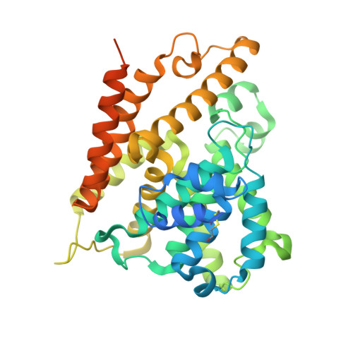
Cyclic nucleotide phosphodiesterases (PDEs) comprise a large family of enzymes that regulate a variety of cellular processes. We describe a family of potent PDE4 inhibitors discovered using an efficient method for scaffold-based drug design. This method involves an iterative approach starting with low-affinity screening of compounds followed by high-throughput cocrystallography to reveal the molecular basis underlying the activity of the newly identified compounds. Through detailed structural analysis of the interaction of the initially discovered pyrazole carboxylic ester scaffold with PDE4D using X-ray crystallography, we identified three sites of chemical substitution and designed small selective libraries of scaffold derivatives with modifications at these sites. A 4,000-fold increase in the potency of this PDE4 inhibitor was achieved after only two rounds of chemical synthesis and the structural analysis of seven pyrazole derivatives bound to PDE4B or PDE4D, revealing the robustness of this approach for identifying new inhibitors that can be further developed into drug candidates.
- Plexxikon Inc., 91 Bolivar Dr., Berkeley, California 94710, USA.
Organizational Affiliation: