X-ray Structures of NADPH-dependent Carbonyl Reductase from Sporobolomyces salmonicolor Provide Insights into Stereoselective Reductions of Carbonyl Compounds
Kamitori, S., Iguchi, A., Ohtaki, A., Yamada, M., Kita, K.(2005) J Mol Biology 352: 551-558
- PubMed: 16095619 Search on PubMed
- DOI: https://doi.org/10.1016/j.jmb.2005.07.011
- Primary Citation Related Structures:
1Y1P, 1ZZE - PubMed Abstract:
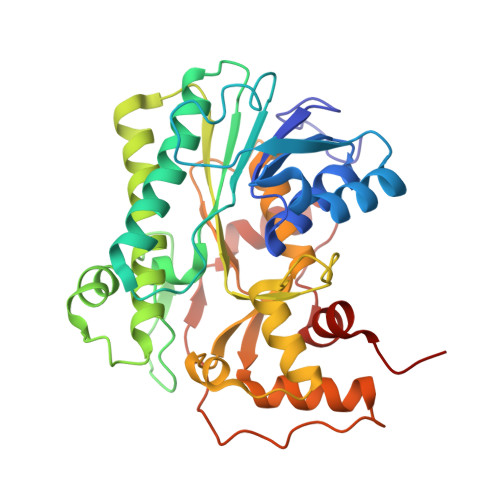
The X-ray structures of red yeast Sporobolomyces salmonicolor carbonyl reductase (SSCR) and its complex with a coenzyme, NADPH, have been determined at a resolution of 1.8A and 1.6A, respectively. SSCR was crystallized in an orthorhombic system with the space group P2(1)2(1)2(1) and cell dimensions of a=54.86 A, b=83.49 A, and c=148.72 A. On its cocrystallization with NADPH, isomorphous crystals of the SSCR/NADPH complex were obtained. The structure of SSCR was solved by a single wavelength anomalous diffraction measurement using a selenomethionine-substituted enzyme, and that of the SSCR/NADPH complex was solved by a molecular replacement method using the solved structure of SSCR. The structures of SSCR and the SSCR/NADPH complex were refined to an R-factor of 0.193 (R(free)=0.233) and 0.211 (R(free)=0.238), respectively. SSCR has two domains, an NADPH-binding domain and a substrate-binding domain, and belongs to the short-chain dehydrogenases/reductases family. The structure of the NADPH-binding domain and the interaction between the enzyme and NADPH are very similar to those found in other structure-solved enzymes belonging to the short-chain dehydrogenases/reductases family, while the structure of the substrate-binding domain is unique. SSCR has stereoselectivity in its catalytic reaction, giving rise to excessive production of (S)-alcohols from ethyl 4-chloro-3-oxobutanoate. The X-ray structure of the SSCR/NADPH complex and preliminary modeling show that the formation of the hydrophobic channel induced by the binding of NADPH is closely related to the stereoselective reduction by SSCR.
- Molecular Structure Research Group, Information Technology Center, Kagawa University, 1750-1 Ikenobe, Miki-cho, Kita-gun, Kagawa 791-0793, Japan. kamitori@med.kagawa-u.ac.jp
Organizational Affiliation: