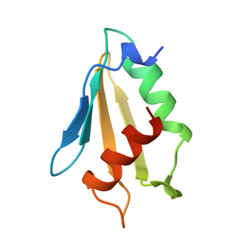
Structure of the oligomerization and L-arginine binding domain of the arginine repressor of Escherichia coli.
Van Duyne, G.D., Ghosh, G., Maas, W.K., Sigler, P.B.(1996) J Mol Biol 256: 377-391
- PubMed: 8594204 Search on PubMed
- DOI: https://doi.org/10.1006/jmbi.1996.0093
- Primary Citation Related Structures:
1XXA, 1XXB, 1XXC - PubMed Abstract:
The structure of the oligomerization and L-arginine binding domain of the Escherichia coli arginine repressor (ArgR) has been determined using X-ray diffraction methods at 2.2 A resolution with bound arginine and at 2.8 A in the unliganded form. The oligomeric core is a 3-fold rotationally symmetric hexamer formed from six identical subunits corresponding to the 77 C-terminal residues (80 to 156) of ArgR. Each subunit has an alpha/beta fold containing a four-stranded antiparallel beta-sheet and two antiparallel alpha-helices. The hexamer is formed from two trimers, each with tightly packed hydrophobic cores. In the absence of arginine, the trimers stack back-to-back through a dyad-symmetric, sparsely packed hydrophobic interface. Six molecules of arginine bind at the trimer-trimer interface, each making ten hydrogen bonds to the protein including a direct ion pair that crosslinks the two protein trimers. Solution experiments with wild-type ArgR and oligomerization domain indicate that the hexameric form is greatly stabilized upon arginine binding. The crystal structures and solution experiments together suggest possible mechanisms of how arginine activates ArgR to bind to its DNA targets and provides a stereochemical basis for interpreting the results of mutagenesis and biochemical experiments with ArgR.
- Department of Molecular Biochemistry and Biophysics, Howard Hughes Medical Institute, Yale University, New Haven, CT 06510 USA.
Organizational Affiliation: