1XN2 | pdb_00001xn2

New substrate binding pockets for beta-secretase.


Experimental Data Snapshot

  • Method: X-RAY DIFFRACTION
  • Resolution: 1.90 Å
  • R-Value Free: 
    0.220 (Depositor) 
  • R-Value Observed: 
    0.193 (Depositor) 

Starting Model: experimental
View more details

wwPDB Validation 3D Report Full Report

Validation slider image for 1XN2

This is version 2.1 of the entry. See complete history

Literature

Structural locations and functional roles of new subsites S5, S6, and S7 in memapsin 2 (beta-secretase).

Turner III, R.T.Hong, L.Koelsch, G.Ghosh, A.K.Tang, J.

(2005) Biochemistry 44: 105-112

  • DOI: https://doi.org/10.1021/bi048106k
  • Primary Citation Related Structures: 
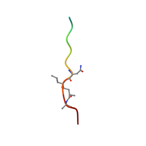
    1XN2, 1XN3

  • PubMed Abstract: 

    Memapsin 2 (beta-secretase) is the membrane-anchored aspartic protease that initiates the cleavage of beta-amyloid precursor protein (APP), leading to the production of amyloid-beta (Abeta), a major factor in the pathogenesis of Alzheimer's disease. The active site of memapsin 2 has been shown, with kinetic data and crystal structures, to bind to eight substrate residues (P(4)-P(4)'). We describe here that the addition of three substrate residues from P(7) to P(5) strongly influences the hydrolytic activity by memapsin 2 and these subsites prefer hydrophobic residues, especially tryptophan. A crystal structure of memapsin 2 complexed with a statine-based inhibitor spanning P(10)-P(4)' revealed the binding positions of P(5)-P(7) residues. Kinetic studies revealed that the addition of these substrate residues contributes to the decrease in K(m) and increase in k(cat) values, suggesting that these residues contribute to both substrate recognition and transition-state binding. The crystal structure of a new inhibitor, OM03-4 (K(i) = 0.03 nM), bound to memapsin 2 revealed the interaction of a tryptophan with the S(6) subsite of the protease.


  • Organizational Affiliation
    • Protein Studies Program, Oklahoma Medical Research Foundation, Department of Biochemistry and Molecular Biology, University of Oklahoma Health Science Center, Oklahoma City, Oklahoma 73104, USA.

Macromolecule Content 

  • Total Structure Weight: 179.81 kDa 
  • Atom Count: 13,323 
  • Modeled Residue Count: 1,596 
  • Deposited Residue Count: 1,604 
  • Unique protein chains: 2

Macromolecules

Find similar proteins by:|  3D Structure
Entity ID: 1
MoleculeChains  Sequence LengthOrganismDetailsImage
Beta-secretase 1
A, B, C, D
389Homo sapiensMutation(s): 0 
Gene Names: BACE1BACE
EC: 3.4.23.46
UniProt & NIH Common Fund Data Resources
Find proteins for P56817 (Homo sapiens)
Explore P56817 
Go to UniProtKB:  P56817
PHAROS:  P56817
GTEx:  ENSG00000186318 
Entity Groups
Sequence Clusters30% Identity50% Identity70% Identity90% Identity95% Identity100% Identity
UniProt GroupP56817
Sequence Annotations
Expand
Reference Sequence
Find similar proteins by:  Sequence   |   3D Structure  
Entity ID: 2
MoleculeChains  Sequence LengthOrganismDetailsImage
OM03-4
E, F, G, H
12N/AMutation(s): 0 

Experimental Data & Validation

Experimental Data

  • Method: X-RAY DIFFRACTION
  • Resolution: 1.90 Å
  • R-Value Free:  0.220 (Depositor) 
  • R-Value Observed: 0.193 (Depositor) 
Space Group: P 1 21 1
Unit Cell:
Length ( Å )Angle ( ˚ )
a = 86.223α = 90
b = 130.784β = 97.71
c = 88.744γ = 90
Software Package:
Software NamePurpose
DENZOdata reduction
SCALEPACKdata scaling
AMoREphasing
CNSrefinement

Structure Validation

View Full Validation Report


Entry History 

Deposition Data

Revision History  (Full details and data files)

  • Version 1.0: 2005-03-22
    Type: Initial release
  • Version 1.1: 2008-04-30
    Changes: Version format compliance
  • Version 1.2: 2011-07-13
    Changes: Atomic model, Database references, Derived calculations, Non-polymer description, Structure summary, Version format compliance
  • Version 1.3: 2023-08-23
    Changes: Data collection, Database references, Derived calculations, Refinement description
  • Version 2.0: 2023-11-15
    Changes: Atomic model, Data collection
  • Version 2.1: 2024-11-13
    Changes: Structure summary