1XKB | pdb_00001xkb

FACTOR XA COMPLEXED WITH A SYNTHETIC INHIBITOR FX-2212A,(2S)-(3'-AMIDINO-3-BIPHENYLYL)-5-(4-PYRIDYLAMINO)PENTANOIC ACID


Experimental Data Snapshot

  • Method: X-RAY DIFFRACTION
  • Resolution: 2.40 Å
  • R-Value Free: 
    0.294 (Depositor) 
  • R-Value Work: 
    0.206 (Depositor) 
  • R-Value Observed: 
    0.206 (Depositor) 

Starting Model: experimental
View more details

wwPDB Validation 3D Report Full Report

Validation slider image for 1XKB

This is version 1.5 of the entry. See complete history

Literature

Structural basis for chemical inhibition of human blood coagulation factor Xa.

Kamata, K.Kawamoto, H.Honma, T.Iwama, T.Kim, S.H.

(1998) Proc Natl Acad Sci U S A 95: 6630-6635

  • DOI: https://doi.org/10.1073/pnas.95.12.6630
  • Primary Citation Related Structures: 
    1XKA, 1XKB

  • PubMed Abstract: 

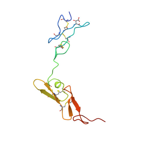
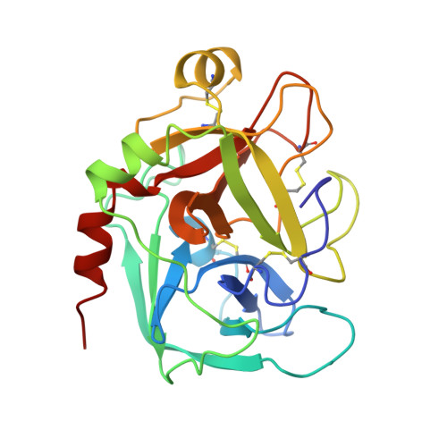
    Factor Xa, the converting enzyme of prothrombin to thrombin, has emerged as an alternative (to thrombin) target for drug discovery for thromboembolic diseases. An inhibitor has been synthesized and the crystal structure of the complex between Des[1-44] factor Xa and the inhibitor has been determined by crystallographic methods in two different crystal forms to 2.3- and 2.4-A resolution. The racemic mixture of inhibitor FX-2212, (2RS)-(3'-amidino-3-biphenylyl)-5-(4-pyridylamino)pentanoic acid, inhibits factor Xa activity by 50% at 272 nM in vitro. The S-isomer of FX-2212 (FX-2212a) was found to bind to the active site of factor Xa in both crystal forms. The biphenylamidine of FX-2212a occupies the S1-pocket, and the pyridine ring makes hydrophobic interactions with the factor Xa aryl-binding site. Several water molecules meditate inhibitor binding to residues in the active site. In contrast to the earlier crystal structures of factor Xa, such as those of apo-Des[1-45] factor Xa and Des[1-44] factor Xa in complex with a naphthyl inhibitor DX-9065a, two epidermal growth factor-like domains of factor Xa are well ordered in both our crystal forms as well as the region between the two domains, which recently was found to be the binding site of the effector cell protease receptor-1. This structure provides a basis for designing next generation inhibitors of factor Xa.


  • Organizational Affiliation
    • Department of Chemistry and Lawrence Berkeley National Laboratory, University of California, Berkeley, CA 94720-5230, USA.

Macromolecule Content 

  • Total Structure Weight: 74.88 kDa 
  • Atom Count: 5,433 
  • Modeled Residue Count: 650 
  • Deposited Residue Count: 660 
  • Unique protein chains: 2

Macromolecules

Find similar proteins by:|  3D Structure
Entity ID: 1
MoleculeChains  Sequence LengthOrganismDetailsImage
BLOOD COAGULATION FACTOR XAA,
C [auth B]
95Homo sapiensMutation(s): 1 
EC: 3.4.21.6
UniProt & NIH Common Fund Data Resources
Find proteins for P00742 (Homo sapiens)
Explore P00742 
Go to UniProtKB:  P00742
PHAROS:  P00742
GTEx:  ENSG00000126218 
Entity Groups
Sequence Clusters30% Identity50% Identity70% Identity90% Identity95% Identity100% Identity
UniProt GroupP00742
Sequence Annotations
Expand
Reference Sequence
Find similar proteins by:|  3D Structure
Entity ID: 2
MoleculeChains  Sequence LengthOrganismDetailsImage
BLOOD COAGULATION FACTOR XAB [auth C],
D
235Homo sapiensMutation(s): 0 
EC: 3.4.21.6
UniProt & NIH Common Fund Data Resources
Find proteins for P00742 (Homo sapiens)
Explore P00742 
Go to UniProtKB:  P00742
PHAROS:  P00742
GTEx:  ENSG00000126218 
Entity Groups
Sequence Clusters30% Identity50% Identity70% Identity90% Identity95% Identity100% Identity
UniProt GroupP00742
Sequence Annotations
Expand
Reference Sequence

Small Molecules

Experimental Data & Validation

Experimental Data

  • Method: X-RAY DIFFRACTION
  • Resolution: 2.40 Å
  • R-Value Free:  0.294 (Depositor) 
  • R-Value Work:  0.206 (Depositor) 
  • R-Value Observed: 0.206 (Depositor) 
Space Group: P 1 21 1
Unit Cell:
Length ( Å )Angle ( ˚ )
a = 58.27α = 90
b = 105.22β = 103.4
c = 63.24γ = 90
Software Package:
Software NamePurpose
DENZOdata reduction
SCALEPACKdata scaling
X-PLORmodel building
X-PLORrefinement
X-PLORphasing

Structure Validation

View Full Validation Report


Entry History 

Deposition Data

Revision History  (Full details and data files)

  • Version 1.0: 1999-03-23
    Type: Initial release
  • Version 1.1: 2008-03-24
    Changes: Version format compliance
  • Version 1.2: 2011-07-13
    Changes: Version format compliance
  • Version 1.3: 2012-06-20
    Changes: Non-polymer description
  • Version 1.4: 2023-08-09
    Changes: Database references, Derived calculations, Refinement description
  • Version 1.5: 2024-11-20
    Changes: Data collection, Structure summary