1XJA

Apo form of the Y31V mutant dimerization domain fragment of Escherichia coli regulatory protein AraC


Experimental Data Snapshot

  • Method: X-RAY DIFFRACTION
  • Resolution: 2.40 Å
  • R-Value Free: 0.256 
  • R-Value Work: 0.211 
  • R-Value Observed: 0.211 

wwPDB Validation   3D Report Full Report


This is version 1.4 of the entry. See complete history


Literature

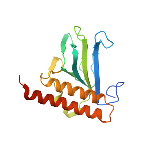
Structure and properties of a truely apo form of AraC dimerization domain.

Weldon, J.E.Rodgers, M.E.Larkin, C.Schleif, R.F.

(2007) Proteins 66: 646-654

  • DOI: https://doi.org/10.1002/prot.21267
  • Primary Citation of Related Structures:  
    1XJA

  • PubMed Abstract: 

    The arabinose-binding pockets of wild type AraC dimerization domains crystallized in the absence of arabinose are occupied with the side chains of Y31 from neighboring domains. This interaction leads to aggregation at high solution concentrations and prevents determination of the structure of truely apo AraC. In this work we found that the aggregation does not significantly occur at physiological concentrations of AraC. We also found that the Y31V mutation eliminates the self-association, but does not affect regulation properties of the protein. At the same time, the mutation allows crystallization of the dimerization domain of the protein with only solvent in the arabinose-binding pocket. Using a distance difference method suitable for detecting and displaying even minor structural variation among large groups of similar structures, we find that there is no significant structural change in the core of monomers of the AraC dimerization domain resulting from arabinose, fucose, or tyrosine occupancy of the ligand-binding pocket. A slight change is observed in the relative orientation of monomers in the dimeric form of the domain upon the binding of arabinose but its significance cannot yet be assessed.


  • Organizational Affiliation

    Department of Biology, Johns Hopkins University, Baltimore, Maryland 21218, USA.


Macromolecules
Find similar proteins by:  (by identity cutoff)  |  3D Structure
Entity ID: 1
MoleculeChains Sequence LengthOrganismDetailsImage
Arabinose operon regulatory protein
A, B, C, D, E
178Escherichia coliMutation(s): 1 
Gene Names: araC
UniProt
Find proteins for P0A9E0 (Escherichia coli (strain K12))
Explore P0A9E0 
Go to UniProtKB:  P0A9E0
Entity Groups  
Sequence Clusters30% Identity50% Identity70% Identity90% Identity95% Identity100% Identity
UniProt GroupP0A9E0
Sequence Annotations
Expand
  • Reference Sequence
Small Molecules
Ligands 1 Unique
IDChains Name / Formula / InChI Key2D Diagram3D Interactions
EDO
Query on EDO

Download Ideal Coordinates CCD File 
AA [auth E]
BA [auth E]
CA [auth E]
DA [auth E]
F [auth A]
AA [auth E],
BA [auth E],
CA [auth E],
DA [auth E],
F [auth A],
G [auth A],
H [auth A],
I [auth B],
J [auth B],
K [auth B],
L [auth B],
M [auth B],
N [auth C],
O [auth C],
P [auth D],
Q [auth D],
R [auth D],
S [auth D],
T [auth D],
U [auth D],
V [auth D],
W [auth D],
X [auth D],
Y [auth D],
Z [auth D]
1,2-ETHANEDIOL
C2 H6 O2
LYCAIKOWRPUZTN-UHFFFAOYSA-N
Experimental Data & Validation

Experimental Data

  • Method: X-RAY DIFFRACTION
  • Resolution: 2.40 Å
  • R-Value Free: 0.256 
  • R-Value Work: 0.211 
  • R-Value Observed: 0.211 
  • Space Group: C 1 2 1
Unit Cell:
Length ( Å )Angle ( ˚ )
a = 260.632α = 90
b = 40.621β = 105.45
c = 102.589γ = 90
Software Package:
Software NamePurpose
CNSrefinement
DENZOdata reduction
SCALEPACKdata scaling
CNSphasing

Structure Validation

View Full Validation Report


Entry History 

Deposition Data

Revision History  (Full details and data files)

  • Version 1.0: 2004-10-12
    Type: Initial release
  • Version 1.1: 2008-04-30
    Changes: Version format compliance
  • Version 1.2: 2011-07-13
    Changes: Version format compliance
  • Version 1.3: 2021-10-20
    Changes: Database references, Derived calculations
  • Version 1.4: 2023-08-23
    Changes: Data collection, Refinement description