Substrate Binding is Required for Assembly of the Active Conformation of the Catalytic Site in Ntn Amidotransferases: Evidence from the 1.8 Angstrom Crystal Structure of the Glutaminase Domain of Glucosamine 6-Phosphate Synthase
Isupov, M.N., Obmolova, G., Butterworth, S., Badet-Denisot, M.-A., Badet, B., Polikarpov, I., Littlechild, J.A., Teplyakov, A.(1996) Structure 4: 801-810
- PubMed: 8805567 Search on PubMed
- DOI: https://doi.org/10.1016/s0969-2126(96)00087-1
- Primary Citation Related Structures:
1XFF, 1XFG - PubMed Abstract:
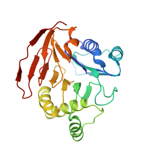
Amidotransferases use the amide nitrogen of glutamine in a number of important biosynthetic reactions. They are composed of a glutaminase domain, which catalyzes the hydrolysis of glutamine to glutamate and ammonia, and a synthetase domain, catalyzing amination of the substrate. To gain insight into the mechanism of nitrogen transfer, we examined the structure of the glutaminase domain of glucosamine 6-phosphate synthase (GLMS). The crystal structures of the enzyme complexed with glutamate and with a competitive inhibitor, Glu-hydroxamate, have been determined to 1.8 A resolution. The protein fold has structural homology to other members of the superfamily of N-terminal nucleophile (Ntn) hydrolases, being a sandwich of antiparallel beta sheets surrounded by two layers of alpha helices. The structural homology between the glutaminase domain of GLMS and that of PRPP amidotransferase (the only other Ntn amidotransferase whose structure is known) indicates that they may have diverged from a common ancestor. Cys1 is the catalytic nucleophile in GLMS, and the nucleophilic character of its thiol group appears to be increased through general base activation by its own alpha-amino group. Cys1 can adopt two conformations, one active and one inactive; glutamine binding locks the residue in a predetermined conformation. We propose that when a nitrogen acceptor is present Cys1 is kept in the active conformation, explaining the phenomenon of substrate-induced activation of the enzyme, and that Arg26 is central in this coupling.
- Department of Chemistry and Biological Sciences, University of Exeter, UK.
Organizational Affiliation: