1XCF

Crystal structure of P28L/Y173F tryptophan synthase alpha-subunits from Escherichia coli


Experimental Data Snapshot

  • Method: X-RAY DIFFRACTION
  • Resolution: 1.80 Å
  • R-Value Free: 0.289 
  • R-Value Work: 0.261 
  • R-Value Observed: 0.261 

wwPDB Validation   3D Report Full Report


This is version 1.4 of the entry. See complete history


Literature

Structures of wild-type and P28L/Y173F tryptophan synthase alpha-subunits from Escherichia coli

Jeong, M.S.Jeong, J.K.Lim, W.K.Jang, S.B.

(2004) Biochem Biophys Res Commun 323: 1257-1264

  • DOI: https://doi.org/10.1016/j.bbrc.2004.08.222
  • Primary Citation of Related Structures:  
    1XC4, 1XCF

  • PubMed Abstract: 

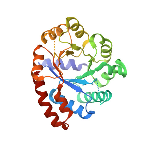
    The alpha-subunit of tryptophan synthase (alphaTS) catalyzes the cleavage of indole-3-glycerol phosphate to glyceraldehyde-3-phosphate and indole, which is used to yield the amino acid tryptophan in tryptophan biosynthesis. Here, we report the first crystal structures of wild-type and double-mutant P28L/Y173F alpha-subunit of tryptophan synthase from Escherichia coli at 2.8 and 1.8A resolution, respectively. The structure of wild-type alphaTS from E. coli was similar to that of the alpha(2)beta(2) complex structure from Salmonella typhimurium. As compared with both structures, the conformational changes are mostly in the interface of alpha- and beta-subunits, and the substrate binding region. Two sulfate ions and two glycerol molecules per asymmetric unit bind with the residues in the active sites of the wild-type structure. Contrarily, double-mutant P28L/Y173F structure is highly closed at the window for the substrate binding by the conformational changes. The P28L substitution induces the exposure of hydrophobic amino acids and decreases the secondary structure that causes the aggregation. The Y173F suppresses to transfer a signal from the alpha-subunit core to the alpha-subunit surface involved in interactions with the beta-subunit and increases structural stability.


  • Organizational Affiliation

    Korea Nanobiotechnology Center, Pusan National University, Jangjeon-dong, Geumjeong-gu, Busan 609-735, Republic of Korea.


Macromolecules
Find similar proteins by:  (by identity cutoff)  |  3D Structure
Entity ID: 1
MoleculeChains Sequence LengthOrganismDetailsImage
Tryptophan synthase alpha chain
A, B
268Escherichia coliMutation(s): 2 
EC: 4.2.1.20
UniProt
Find proteins for P0A877 (Escherichia coli (strain K12))
Explore P0A877 
Go to UniProtKB:  P0A877
Entity Groups  
Sequence Clusters30% Identity50% Identity70% Identity90% Identity95% Identity100% Identity
UniProt GroupP0A877
Sequence Annotations
Expand
  • Reference Sequence
Experimental Data & Validation

Experimental Data

  • Method: X-RAY DIFFRACTION
  • Resolution: 1.80 Å
  • R-Value Free: 0.289 
  • R-Value Work: 0.261 
  • R-Value Observed: 0.261 
  • Space Group: P 1 21 1
Unit Cell:
Length ( Å )Angle ( ˚ )
a = 71.087α = 90
b = 52.695β = 91.48
c = 71.518γ = 90
Software Package:
Software NamePurpose
DENZOdata reduction
SCALEPACKdata scaling
AMoREphasing
CNSrefinement

Structure Validation

View Full Validation Report


Entry History 

Deposition Data

  • Released Date: 2004-11-02 
  • Deposition Author(s): Jang, S.B.

Revision History  (Full details and data files)

  • Version 1.0: 2004-11-02
    Type: Initial release
  • Version 1.1: 2008-04-30
    Changes: Version format compliance
  • Version 1.2: 2011-07-13
    Changes: Version format compliance
  • Version 1.3: 2021-11-10
    Changes: Database references
  • Version 1.4: 2023-10-25
    Changes: Data collection, Refinement description