1XAP

Structure of the ligand binding domain of the Retinoic Acid Receptor beta


Experimental Data Snapshot

  • Method: X-RAY DIFFRACTION
  • Resolution: 2.10 Å
  • R-Value Free: 0.253 
  • R-Value Work: 0.213 
  • R-Value Observed: 0.213 

wwPDB Validation   3D Report Full Report


Ligand Structure Quality Assessment 


This is version 1.4 of the entry. See complete history


Literature

Rational design of RAR-selective ligands revealed by RARbeta crystal structure

Germain, P.Kammerer, S.Perez, E.Peluso-Iltis, C.Tortolani, D.Zusi, F.C.Starrett, J.Lapointe, P.Daris, J.P.Marinier, A.De Lera, A.R.Rochel, N.Gronemeyer, H.

(2004) EMBO Rep 5: 877-882

  • DOI: https://doi.org/10.1038/sj.embor.7400235
  • Primary Citation of Related Structures:  
    1XAP

  • PubMed Abstract: 

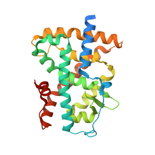
    The crystal structure of the ligand-binding domain of RARbeta, a suspect tumour suppressor, reveals important features that distinguish it from the two other RAR isotypes. The most striking difference is an extra cavity allowing RARbeta to bind more bulky agonists. Accordingly, we identified a ligand that shows RARbeta selectivity with a 100-fold higher affinity to RARbeta than to alpha or gamma isotypes. The structural differences between the three RAR ligand-binding pockets revealed a rationale explaining how a single retinoid can be at the same time an RARalpha, gamma antagonist and an RARbeta agonist. In addition, we demonstrate how to generate an RARbeta antagonist by gradually modifying the bulkiness of a single substitution. Together, our results provide structural guidelines for the synthesis of RARbeta-selective agonists and antagonists, allowing for the first time to address pharmacologically the tumour suppressor role of RARbeta in vitro and in animal models.


  • Organizational Affiliation

    Institut de Génétique et de Biologie Moléculaire et Cellulaire CNRS/INSERM/ULP, BP 10142, 67404 Illkirch Cedex, CU de Strasbourg, France.


Macromolecules
Find similar proteins by:  (by identity cutoff)  |  3D Structure
Entity ID: 1
MoleculeChains Sequence LengthOrganismDetailsImage
Retinoic acid receptor beta267Homo sapiensMutation(s): 0 
UniProt & NIH Common Fund Data Resources
Find proteins for P10826 (Homo sapiens)
Explore P10826 
Go to UniProtKB:  P10826
PHAROS:  P10826
GTEx:  ENSG00000077092 
Entity Groups  
Sequence Clusters30% Identity50% Identity70% Identity90% Identity95% Identity100% Identity
UniProt GroupP10826
Sequence Annotations
Expand
  • Reference Sequence
Small Molecules
Ligands 1 Unique
IDChains Name / Formula / InChI Key2D Diagram3D Interactions
TTB
Query on TTB

Download Ideal Coordinates CCD File 
B [auth A]4-[(1E)-2-(5,5,8,8-TETRAMETHYL-5,6,7,8-TETRAHYDRONAPHTHALEN-2-YL)PROP-1-ENYL]BENZOIC ACID
C24 H28 O2
FOIVPCKZDPCJJY-JQIJEIRASA-N
Binding Affinity Annotations 
IDSourceBinding Affinity
TTB BindingDB:  1XAP Ki: min: 0.5, max: 21 (nM) from 4 assay(s)
Kd: min: 5, max: 36 (nM) from 3 assay(s)
IC50: min: 5, max: 36 (nM) from 3 assay(s)
EC50: min: 0.06, max: 85 (nM) from 13 assay(s)
PDBBind:  1XAP Kd: 2 (nM) from 1 assay(s)
Binding MOAD:  1XAP Kd: 2.5 (nM) from 1 assay(s)
Experimental Data & Validation

Experimental Data

  • Method: X-RAY DIFFRACTION
  • Resolution: 2.10 Å
  • R-Value Free: 0.253 
  • R-Value Work: 0.213 
  • R-Value Observed: 0.213 
  • Space Group: P 21 21 21
Unit Cell:
Length ( Å )Angle ( ˚ )
a = 47.276α = 90
b = 57.808β = 90
c = 90.289γ = 90
Software Package:
Software NamePurpose
HKL-2000data collection
SCALEPACKdata scaling
CNSrefinement
HKL-2000data reduction
CNSphasing

Structure Validation

View Full Validation Report


Ligand Structure Quality Assessment 


Entry History 

Revision History  (Full details and data files)

  • Version 1.0: 2004-11-16
    Type: Initial release
  • Version 1.1: 2008-04-30
    Changes: Version format compliance
  • Version 1.2: 2011-07-13
    Changes: Version format compliance
  • Version 1.3: 2016-12-28
    Changes: Database references
  • Version 1.4: 2023-10-25
    Changes: Data collection, Database references, Derived calculations, Refinement description