The structures of the unique sulfotransferase retinol dehydratase with product and inhibitors provide insight into enzyme mechanism and inhibition.
Pakhomova, S., Buck, J., Newcomer, M.E.(2005) Protein Sci 14: 176-182
- PubMed: 15608121 Search on PubMedSearch on PubMed Central
- DOI: https://doi.org/10.1110/ps.041061105
- Primary Citation Related Structures:
1X8J, 1X8K, 1X8L - PubMed Abstract:
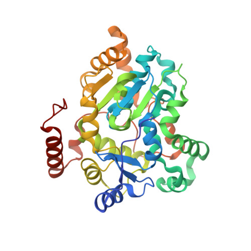
The structure of retinol dehydratase (DHR) from Spodoptera frugiperda, a member of the sulfotransferase superfamily, in complexes with the inactive form of the cofactor PAP 3'-phosphoadenosine 5'-phosphate (PAP) and (1) the product of the reaction with retinol anhydroretinol (AR), (2) the retinoid inhibitor all-trans-4-oxoretinol (OR), and (3) the potent steroid inhibitor androsterone (AND) have been determined and compared to the enzyme complex with PAP and retinol. The structures show that the geometry of the active-site amino acids is largely preserved in the various complexes. However, the beta-ionone rings of the retinoids are oriented differently with respect to side chains that have been shown to be important for the enzymatic reaction. In addition, the DHR:PAP:AND complex reveals a novel mode for steroid binding that contrasts significantly with that for steroid binding in other sulfotransferases. The molecule is displaced and rotated approximately 180 degrees along its length so that there is no acceptor hydroxyl in close proximity to the site of sulfate transfer. This observation explains why steroids are potent inhibitors of retinol dehydratase activity, rather than substrates for sulfonation. Most of the steroid-protein contacts are provided by the alpha-helical cap that distinguishes this member of the superfamily. This observation suggests that in addition to providing a chemical environment that promotes the dehydration of a sulfonated intermediate, the cap may also serve to minimize a promiscuous sulfotransferases activity.
- Department of Biological Sciences, Louisiana State University, Baton Rouge, LA 70803, USA.
Organizational Affiliation: