1X2H | pdb_00001x2h

Crystal Structure of Lipate-Protein Ligase A from Escherichia coli complexed with lipoic acid


Experimental Data Snapshot

  • Method: X-RAY DIFFRACTION
  • Resolution: 2.91 Å
  • R-Value Free: 
    0.271 (Depositor), 0.266 (DCC) 
  • R-Value Work: 
    0.186 (Depositor), 0.184 (DCC) 
  • R-Value Observed: 
    0.191 (Depositor) 

Starting Model: experimental
View more details

wwPDB Validation 3D Report Full Report

Validation slider image for 1X2H

Ligand Structure Quality Assessment 


This is version 1.5 of the entry. See complete history

Literature

Crystal structure of lipoate-protein ligase A from Escherichia coli: Determination of the lipoic acid-binding site

Fujiwara, K.Toma, S.Okamura-Ikeda, K.Motokawa, Y.Nakagawa, A.Taniguchi, H.

(2005) J Biological Chem 280: 33645-33651

  • DOI: https://doi.org/10.1074/jbc.M505010200
  • Primary Citation Related Structures: 
    1X2G, 1X2H

  • PubMed Abstract: 

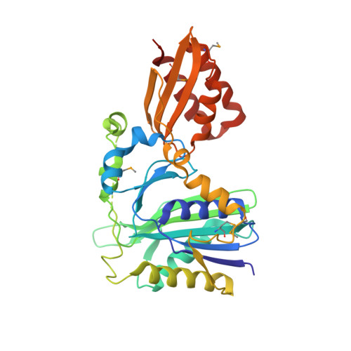
    Lipoate-protein ligase A (LplA) catalyzes the formation of lipoyl-AMP from lipoate and ATP and then transfers the lipoyl moiety to a specific lysine residue on the acyltransferase subunit of alpha-ketoacid dehydrogenase complexes and on H-protein of the glycine cleavage system. The lypoyllysine arm plays a pivotal role in the complexes by shuttling the reaction intermediate and reducing equivalents between the active sites of the components of the complexes. We have determined the X-ray crystal structures of Escherichia coli LplA alone and in a complex with lipoic acid at 2.4 and 2.9 angstroms resolution, respectively. The structure of LplA consists of a large N-terminal domain and a small C-terminal domain. The structure identifies the substrate binding pocket at the interface between the two domains. Lipoic acid is bound in a hydrophobic cavity in the N-terminal domain through hydrophobic interactions and a weak hydrogen bond between carboxyl group of lipoic acid and the Ser-72 or Arg-140 residue of LplA. No large conformational change was observed in the main chain structure upon the binding of lipoic acid.


  • Organizational Affiliation
    • Institute for Enzyme Research, the University of Tokushima, Tokushima 770-8503, Japan. fujiwara@ier.tokushima-u.ac.jp

Macromolecule Content 

  • Total Structure Weight: 114.64 kDa 
  • Atom Count: 7,937 
  • Modeled Residue Count: 997 
  • Deposited Residue Count: 1,011 
  • Unique protein chains: 1

Macromolecules

Find similar proteins by:|  3D Structure
Entity ID: 1
MoleculeChains  Sequence LengthOrganismDetailsImage
Lipoate-protein ligase A
A, B, C
337Escherichia coliMutation(s): 5 
EC: 6.3.2 (PDB Primary Data), 6.3.1.20 (UniProt)
UniProt
Find proteins for P32099 (Escherichia coli (strain K12))
Explore P32099 
Go to UniProtKB:  P32099
Entity Groups
Sequence Clusters30% Identity50% Identity70% Identity90% Identity95% Identity100% Identity
UniProt GroupP32099
Sequence Annotations
Expand
Reference Sequence

Small Molecules

Modified Residues  1 Unique
IDChains TypeFormula2D DiagramParent
MSE
Query on MSE
A, B, C
L-PEPTIDE LINKINGC5 H11 N O2 SeMET

Experimental Data & Validation

Experimental Data

  • Method: X-RAY DIFFRACTION
  • Resolution: 2.91 Å
  • R-Value Free:  0.271 (Depositor), 0.266 (DCC) 
  • R-Value Work:  0.186 (Depositor), 0.184 (DCC) 
  • R-Value Observed: 0.191 (Depositor) 
Space Group: C 2 2 21
Unit Cell:
Length ( Å )Angle ( ˚ )
a = 83.2α = 90
b = 111.6β = 90
c = 289.6γ = 90
Software Package:
Software NamePurpose
REFMACrefinement
HKL-2000data reduction
SCALEPACKdata scaling

Structure Validation

View Full Validation Report


Ligand Structure Quality Assessment 


Entry History 

Deposition Data

Revision History  (Full details and data files)

  • Version 1.0: 2005-08-02
    Type: Initial release
  • Version 1.1: 2008-04-30
    Changes: Version format compliance
  • Version 1.2: 2011-07-13
    Changes: Version format compliance
  • Version 1.3: 2017-10-11
    Changes: Refinement description
  • Version 1.4: 2024-04-03
    Changes: Data collection, Database references, Derived calculations, Refinement description
  • Version 1.5: 2024-10-16
    Changes: Structure summary