


Structure of 6-aminohexanoate-dimer hydrolase
Negoro, S., Ohki, T., Shibata, N., Mizuno, N., Wakitani, Y., Tsurukame, J., Matsumoto, K., Kawamoto, I., Takeo, M., Higuchi, Y.To be published.
Experimental Data Snapshot
wwPDB Validation 3D Report Full Report

Entity ID: 1 | |||||
|---|---|---|---|---|---|
| Molecule | Chains | Sequence Length | Organism | Details | Image |
| 6-aminohexanoate-dimer hydrolase | 392 | Flavobacterium sp. | Mutation(s): 0 EC: 3.5.1.46 | 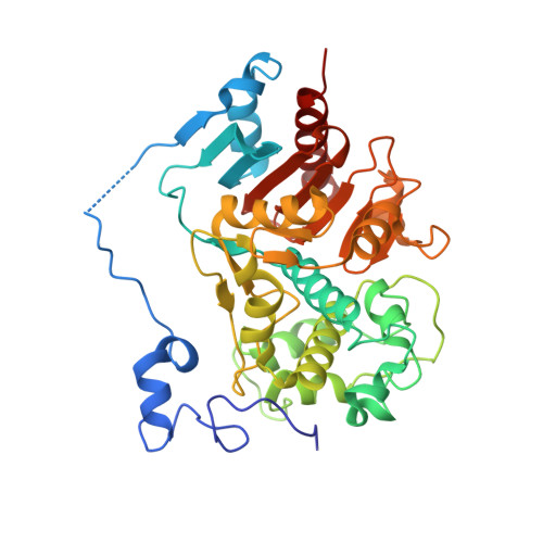 | |
UniProt | |||||
Entity Groups | |||||
| Sequence Clusters | 30% Identity50% Identity70% Identity90% Identity95% Identity100% Identity | ||||
| UniProt Group | P07062 | ||||
Sequence AnnotationsExpand | |||||
Reference Sequence | |||||
| Length ( Å ) | Angle ( ˚ ) |
|---|---|
| a = 96.37 | α = 90 |
| b = 96.37 | β = 90 |
| c = 113.09 | γ = 120 |
| Software Name | Purpose |
|---|---|
| CNS | refinement |
| MOSFLM | data reduction |
| CCP4 | data scaling |
| SHARP | phasing |


