1WS5

Crystal structure of Jacalin-Me-alpha-Mannose complex: Promiscuity vs Specificity


Experimental Data Snapshot

  • Method: X-RAY DIFFRACTION
  • Resolution: 1.90 Å
  • R-Value Free: 0.204 
  • R-Value Work: 0.185 
  • R-Value Observed: 0.185 

wwPDB Validation   3D Report Full Report


Ligand Structure Quality Assessment 


This is version 1.5 of the entry. See complete history


Literature

Structural basis for the energetics of jacalin-sugar interactions: promiscuity versus specificity

Jeyaprakash, A.A.Jayashree, G.Mahanta, S.K.Swaminathan, C.P.Sekar, K.Surolia, A.Vijayan, M.

(2005) J Mol Biol 347: 181-188

  • DOI: https://doi.org/10.1016/j.jmb.2005.01.015
  • Primary Citation of Related Structures:  
    1WS4, 1WS5

  • PubMed Abstract: 

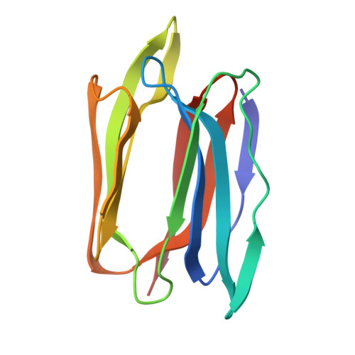
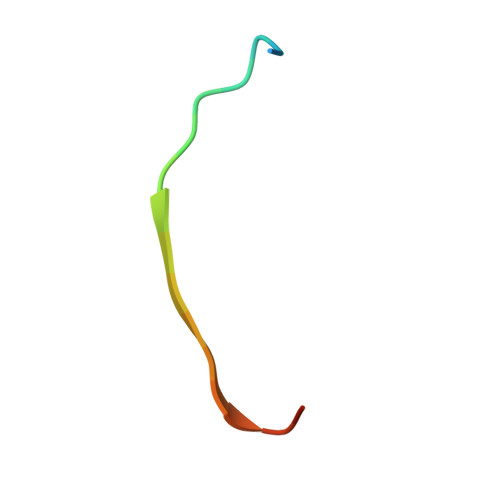
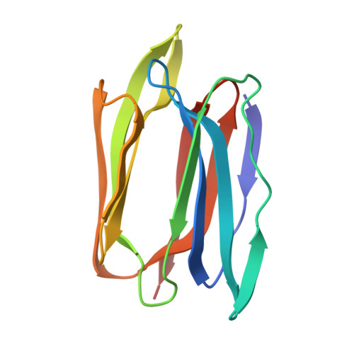
    Jacalin, a tetrameric lectin, is one of the two lectins present in jackfruit (Artocarpus integrifolia) seeds. Its crystal structure revealed, for the first time, the occurrence of the beta-prism I fold in lectins. The structure led to the elucidation of the crucial role of a new N terminus generated by post-translational proteolysis for the lectin's specificity for galactose. Subsequent X-ray studies on other carbohydrate complexes showed that the extended binding site of jacalin consisted of, in addition to the primary binding site, a hydrophobic secondary site A composed of aromatic residues and a secondary site B involved mainly in water-bridges. A recent investigation involving surface plasmon resonance and the X-ray analysis of a methyl-alpha-mannose complex, had led to a suggestion of promiscuity in the lectin's sugar specificity. To explore this suggestion further, detailed isothermal titration calorimetric studies on the interaction of galactose (Gal), mannose (Man), glucose (Glc), Me-alpha-Gal, Me-alpha-Man, Me-alpha-Glc and other mono- and oligosaccharides of biological relevance and crystallographic studies on the jacalin-Me-alpha-Glc complex and a new form of the jacalin-Me-alpha-Man complex, have been carried out. The binding affinity of Me-alpha-Man is 20 times weaker than that of Me-alpha-Gal. The corresponding number is 27, when the binding affinities of Gal and Me-alpha-Gal, and those of Man and Me-alpha-Man are compared. Glucose (Glc) shows no measurable binding, while the binding affinity of Me-alpha-Glc is slightly less than that of Me-alpha-Man. The available crystal structures of jacalin-sugar complexes provide a convincing explanation for the energetics of binding in terms of interactions at the primary binding site and secondary site A. The other sugars used in calorimetric studies show no detectable binding to jacalin. These results and other available evidence suggest that jacalin is specific to O-glycans and its affinity to N-glycans is extremely weak or non-existent and therefore of limited value in processes involving biological recognition.


  • Organizational Affiliation

    Molecular Biophysics Unit, Indian Institute of Science, Bangalore 560 012, India.


Macromolecules
Find similar proteins by:  (by identity cutoff)  |  3D Structure
Entity ID: 1
MoleculeChains Sequence LengthOrganismDetailsImage
Agglutinin alpha chain
A, G
133Artocarpus integerMutation(s): 0 
UniProt
Find proteins for P18670 (Artocarpus integer)
Explore P18670 
Go to UniProtKB:  P18670
Entity Groups  
Sequence Clusters30% Identity50% Identity70% Identity90% Identity95% Identity100% Identity
UniProt GroupP18670
Sequence Annotations
Expand
  • Reference Sequence

Find similar proteins by:  Sequence   |   3D Structure  

Entity ID: 2
MoleculeChains Sequence LengthOrganismDetailsImage
Agglutinin beta-3 chain
B, D, F, H
20Artocarpus integerMutation(s): 0 
UniProt
Find proteins for P18673 (Artocarpus integer)
Explore P18673 
Go to UniProtKB:  P18673
Entity Groups  
Sequence Clusters30% Identity50% Identity70% Identity90% Identity95% Identity100% Identity
UniProt GroupP18673
Sequence Annotations
Expand
  • Reference Sequence
Find similar proteins by:  (by identity cutoff)  |  3D Structure
Entity ID: 3
MoleculeChains Sequence LengthOrganismDetailsImage
Agglutinin alpha chain
C, E
133Artocarpus integerMutation(s): 0 
UniProt
Find proteins for P18670 (Artocarpus integer)
Explore P18670 
Go to UniProtKB:  P18670
Entity Groups  
Sequence Clusters30% Identity50% Identity70% Identity90% Identity95% Identity100% Identity
UniProt GroupP18670
Sequence Annotations
Expand
  • Reference Sequence
Small Molecules
Experimental Data & Validation

Experimental Data

  • Method: X-RAY DIFFRACTION
  • Resolution: 1.90 Å
  • R-Value Free: 0.204 
  • R-Value Work: 0.185 
  • R-Value Observed: 0.185 
  • Space Group: P 21 21 21
Unit Cell:
Length ( Å )Angle ( ˚ )
a = 78.477α = 90
b = 99.097β = 90
c = 105.659γ = 90
Software Package:
Software NamePurpose
CNSrefinement
MAR345data collection
SCALEPACKdata scaling
CNSphasing

Structure Validation

View Full Validation Report


Ligand Structure Quality Assessment 


Entry History 

Deposition Data

Revision History  (Full details and data files)

  • Version 1.0: 2005-03-29
    Type: Initial release
  • Version 1.1: 2008-04-30
    Changes: Version format compliance
  • Version 1.2: 2011-07-13
    Changes: Version format compliance
  • Version 1.3: 2017-10-11
    Changes: Refinement description
  • Version 1.4: 2020-07-29
    Type: Remediation
    Reason: Carbohydrate remediation
    Changes: Data collection, Database references, Derived calculations, Structure summary
  • Version 1.5: 2023-10-25
    Changes: Data collection, Database references, Refinement description, Structure summary