Biochemical and crystallographic analyses of maltohexaose-producing amylase from alkalophilic Bacillus sp. 707
Kanai, R., Haga, K., Akiba, T., Yamane, K., Harata, K.(2004) Biochemistry 43: 14047-14056
- PubMed: 15518553 Search on PubMed
- DOI: https://doi.org/10.1021/bi048489m
- Primary Citation Related Structures:
1WP6, 1WPC - PubMed Abstract:
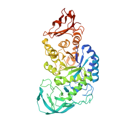
Maltohexaose-producing amylase, called G6-amylase (EC 3.2.1.98), from alkalophilic Bacillus sp.707 predominantly produces maltohexaose (G6) from starch and related alpha-1,4-glucans. To elucidate the reaction mechanism of G6-amylase, the enzyme activities were evaluated and crystal structures were determined for the native enzyme and its complex with pseudo-maltononaose at 2.1 and 1.9 A resolutions, respectively. The optimal condition for starch-degrading reaction activity was found at 45 degrees C and pH 8.8, and the enzyme produced G6 in a yield of more than 30% of the total products from short-chain amylose (DP = 17). The crystal structures revealed that Asp236 is a nucleophilic catalyst and Glu266 is a proton donor/acceptor. Pseudo-maltononaose occupies subsites -6 to +3 and induces the conformational change of Glu266 and Asp333 to form a salt linkage with the N-glycosidic amino group and a hydrogen bond with secondary hydroxyl groups of the cyclitol residue bound to subsite -1, respectively. The indole moiety of Trp140 is stacked on the cyclitol and 4-amino-6-deoxyglucose residues located at subsites -6 and -5 within a 4 A distance. Such a face-to-face short contact may regulate the disposition of the glucosyl residue at subsite -6 and would govern the product specificity for G6 production.
- Institute of Biological Sciences, University of Tsukuba, Tsukuba, Ibaraki 305-8572, Japan.
Organizational Affiliation: