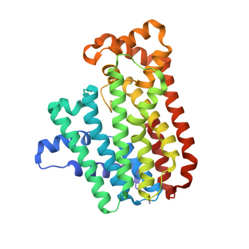
Crystal structure of geranulgeranyl diphosphate synthase from Thermus thermophilus
Suto, K., Nishio, K., Nodake, Y., Hamada, K., Kawamoto, M., Nakagawa, N., Kuramitu, S., Miura, K.To be published.
Experimental Data Snapshot
wwPDB Validation 3D Report Full Report
Entity ID: 1 | |||||
|---|---|---|---|---|---|
| Molecule | Chains | Sequence Length | Organism | Details | Image |
| geranylgeranyl diphosphate synthetase | 330 | Thermus thermophilus HB8 | Mutation(s): 0 EC: 2.5.1 |  | |
UniProt | |||||
Find proteins for Q5SMD0 (Thermus thermophilus (strain ATCC 27634 / DSM 579 / HB8)) Explore Q5SMD0 Go to UniProtKB: Q5SMD0 | |||||
Entity Groups | |||||
| Sequence Clusters | 30% Identity50% Identity70% Identity90% Identity95% Identity100% Identity | ||||
| UniProt Group | Q5SMD0 | ||||
Sequence AnnotationsExpand | |||||
| |||||
| Length ( Å ) | Angle ( ˚ ) |
|---|---|
| a = 139.88 | α = 90 |
| b = 139.88 | β = 90 |
| c = 73.35 | γ = 90 |
| Software Name | Purpose |
|---|---|
| CNS | refinement |
| HKL-2000 | data reduction |
| SCALEPACK | data scaling |
| SOLVE | phasing |