1WBX | pdb_00001wbx

CRYSTAL STRUCTURES OF MURINE MHC CLASS I H-2 Db AND Kb MOLECULES IN COMPLEX WITH CTL EPITOPES FROM INFLUENZA A VIRUS: IMPLICATIONS FOR TCR REPERTOIRE SELECTION AND IMMUNODOMINANCE


Experimental Data Snapshot

  • Method: X-RAY DIFFRACTION
  • Resolution: 1.90 Å
  • R-Value Free: 
    0.255 (Depositor), 0.262 (DCC) 
  • R-Value Work: 
    0.198 (Depositor), 0.203 (DCC) 
  • R-Value Observed: 
    0.204 (Depositor) 

Starting Model: experimental
View more details

wwPDB Validation 3D Report Full Report

Validation slider image for 1WBX

This is version 1.4 of the entry. See complete history

Literature

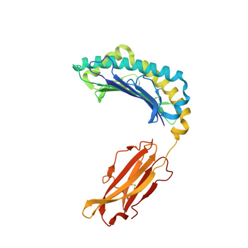
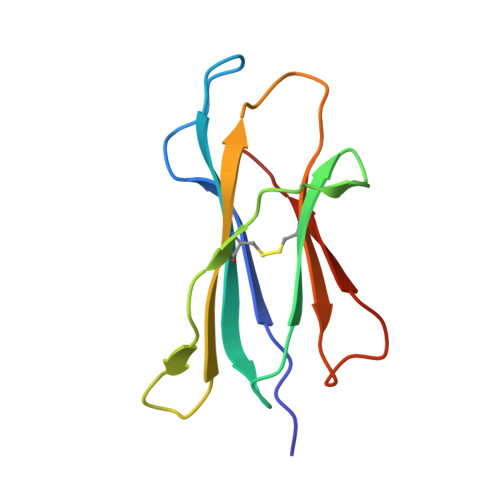
Crystal Structures of Murine Mhc Class I H-2 D(B) and K(B) Molecules in Complex with Ctl Epitopes from Influenza a Virus: Implications for Tcr Repertoire Selection and Immunodominance

Meijers, R.Lai, C.Yang, Y.Liu, J.Zhong, W.Wang, J.Reinherz, E.L.

(2005) J Mol Biology 345: 1099

  • DOI: https://doi.org/10.1016/j.jmb.2004.11.023
  • Primary Citation Related Structures: 
    1WBX, 1WBY, 1WBZ

  • PubMed Abstract: 

    Cytotoxic T lymphocyte (CTL) responses against influenza A virus in C57BL/6 mice are dominated by a small number of viral peptides among many that are capable of binding to major histocompatibility complex (MHC) class I molecules. The basis of this limited immune recognition is unknown. Here, we present X-ray structures of MHC class I molecules in complex with two immunodominant epitopes (PA(224-233)/D(b) and PB1(703-711)/K(b)) and one non-immunogenic epitope (HA(468-477)/D(b)) of the influenza A virus. The immunodominant peptides are each characterized by a bulge at the C terminus, lifting P6 and P7 residues out of the MHC groove, presenting featured structural elements to T-cell receptors (TCRs). Immune recognition of PA(224-233)/D(b) will focus largely on the exposed P7 arginine residue. In contrast, the non-immunogenic HA(468-477) peptide lacks prominent features in this C-terminal bulge. In the K(b)-bound PB1(703-711) epitope, the bulge results from a non-canonical binding motif, such that the mode of presentation of this peptide strongly resembles that of D(b)-bound peptides. Given that PA(224-233)/D(b), PB1(703-711)/K(b) and the previously defined NP(366-374)/D(b) epitopes dominate the primary response to influenza A virus in C57BL/6 mice, our findings indicate that residues of the C-terminal bulge are important in selection of the immunodominant CTL repertoire.


  • Organizational Affiliation
    • Laboratory of Immunobiology, Department of Medical Oncology, Dana-Farber Cancer Institute, Harvard Medical School, Boston, MA 02115, USA.

Macromolecule Content 

  • Total Structure Weight: 44.94 kDa 
  • Atom Count: 3,677 
  • Modeled Residue Count: 384 
  • Deposited Residue Count: 385 
  • Unique protein chains: 3

Macromolecules

Find similar proteins by:|  3D Structure
Entity ID: 1
MoleculeChains  Sequence LengthOrganismDetailsImage
H-2 CLASS I HISTOCOMPATIBILITY ANTIGEN, D-B ALPHA CHAIN276Mus musculusMutation(s): 0 
UniProt
Find proteins for P01899 (Mus musculus)
Explore P01899 
Go to UniProtKB:  P01899
Entity Groups
Sequence Clusters30% Identity50% Identity70% Identity90% Identity95% Identity100% Identity
UniProt GroupP01899
Sequence Annotations
Expand
Reference Sequence
Find similar proteins by:|  3D Structure
Entity ID: 2
MoleculeChains  Sequence LengthOrganismDetailsImage
BETA-2MICROGLOBULIN99Mus musculusMutation(s): 0 
UniProt & NIH Common Fund Data Resources
Find proteins for P01887 (Mus musculus)
Explore P01887 
Go to UniProtKB:  P01887
IMPC:  MGI:88127
Entity Groups
Sequence Clusters30% Identity50% Identity70% Identity90% Identity95% Identity100% Identity
UniProt GroupP01887
Sequence Annotations
Expand
Reference Sequence
Find similar proteins by:  Sequence   |   3D Structure  
Entity ID: 3
MoleculeChains  Sequence LengthOrganismDetailsImage
INFLUENZA A PEPTIDE10Influenza A virusMutation(s): 0 
UniProt
Find proteins for P26140 (Influenza A virus (strain A/Swine/Indiana/1726/1988 H1N1))
Explore P26140 
Go to UniProtKB:  P26140
Entity Groups
Sequence Clusters30% Identity50% Identity70% Identity90% Identity95% Identity100% Identity
UniProt GroupP26140
Sequence Annotations
Expand
Reference Sequence

Experimental Data & Validation

Experimental Data

  • Method: X-RAY DIFFRACTION
  • Resolution: 1.90 Å
  • R-Value Free:  0.255 (Depositor), 0.262 (DCC) 
  • R-Value Work:  0.198 (Depositor), 0.203 (DCC) 
  • R-Value Observed: 0.204 (Depositor) 
Space Group: P 43 21 2
Unit Cell:
Length ( Å )Angle ( ˚ )
a = 56.759α = 90
b = 56.759β = 90
c = 275.996γ = 90
Software Package:
Software NamePurpose
REFMACrefinement
SCALEPACKdata scaling
AMoREphasing

Structure Validation

View Full Validation Report


Entry History 

Deposition Data

Revision History  (Full details and data files)

  • Version 1.0: 2005-01-19
    Type: Initial release
  • Version 1.1: 2011-05-08
    Changes: Version format compliance
  • Version 1.2: 2011-07-13
    Changes: Version format compliance
  • Version 1.3: 2023-12-13
    Changes: Data collection, Database references, Other, Refinement description
  • Version 1.4: 2024-11-20
    Changes: Structure summary