1WBF | pdb_00001wbf

WINGED BEAN LECTIN, SACCHARIDE FREE FORM


Experimental Data Snapshot

  • Method: X-RAY DIFFRACTION
  • Resolution: 2.30 Å
  • R-Value Free: 
    0.257 (Depositor) 
  • R-Value Work: 
    0.197 (Depositor), 0.198 (DCC) 

Starting Model: experimental
View more details

wwPDB Validation 3D Report Full Report

Validation slider image for 1WBF

Ligand Structure Quality Assessment 


This is version 1.5 of the entry. See complete history

Literature

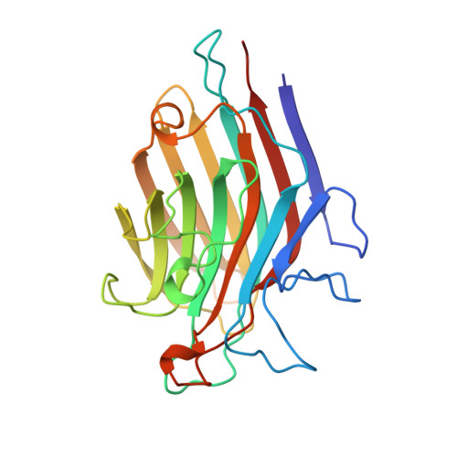
Structure of basic winged-bean lectin and a comparison with its saccharide-bound form.

Manoj, N.Srinivas, V.R.Suguna, K.

(1999) Acta Crystallogr D Biol Crystallogr 55: 794-800

  • DOI: https://doi.org/10.1107/s090744499900044x
  • Primary Citation Related Structures: 
    1WBF

  • PubMed Abstract: 

    The crystal structure of the saccharide-free form of the basic form of winged-bean agglutinin (WBAI) has been solved by the molecular-replacement method and refined at 2.3 A resolution. The final R factor is 19.7% for all data in the resolution range 8.0-2.3 A. The asymmetric unit contains two half-dimers, each located on a crystallographic twofold axis. The structure of the saccharide-free form is compared with that of the complex of WBAI with methyl-alpha-D-galactoside. The complex is composed of two dimers in the asymmetric unit. The intersubunit interactions in the dimer are nearly identical in the two structures. The binding site of the saccharide-free structure contains three ordered water molecules at positions similar to those of the hydroxyl groups of the carbohydrate which are hydrogen bonded to the protein. Superposition of the saccharide-binding sites of the two structures shows that the major changes involve expulsion of these ordered water molecules and a shift of about 0.6 A of the main-chain atoms of the variable loop.


  • Organizational Affiliation
    • Molecular Biophysics Unit, Indian Institute of Science, Bangalore-560 012, India.

Macromolecule Content 

  • Total Structure Weight: 54.35 kDa 
  • Atom Count: 3,965 
  • Modeled Residue Count: 474 
  • Deposited Residue Count: 484 
  • Unique protein chains: 1

Macromolecules

Find similar proteins by:|  3D Structure
Entity ID: 1
MoleculeChains  Sequence LengthOrganismDetailsImage
PROTEIN (AGGLUTININ)
A, B
242Psophocarpus tetragonolobusMutation(s): 0 
UniProt
Find proteins for O24313 (Psophocarpus tetragonolobus)
Explore O24313 
Go to UniProtKB:  O24313
Entity Groups
Sequence Clusters30% Identity50% Identity70% Identity90% Identity95% Identity100% Identity
UniProt GroupO24313
Glycosylation
Glycosylation Sites: 2
Sequence Annotations
Expand
Reference Sequence

Small Molecules

Ligands 3 Unique
IDChains Name / Formula / InChI Key2D Diagram3D Interactions
NAG

Query on NAG


Download:Ideal Coordinates CCD File
C [auth A],
D [auth A],
G [auth B],
H [auth B]
2-acetamido-2-deoxy-beta-D-glucopyranose
C8 H15 N O6
OVRNDRQMDRJTHS-FMDGEEDCSA-N
MN

Query on MN


Download:Ideal Coordinates CCD File
E [auth A],
I [auth B]
MANGANESE (II) ION
Mn
WAEMQWOKJMHJLA-UHFFFAOYSA-N
CA

Query on CA


Download:Ideal Coordinates CCD File
F [auth A],
J [auth B]
CALCIUM ION
Ca
BHPQYMZQTOCNFJ-UHFFFAOYSA-N

Experimental Data & Validation

Experimental Data

  • Method: X-RAY DIFFRACTION
  • Resolution: 2.30 Å
  • R-Value Free:  0.257 (Depositor) 
  • R-Value Work:  0.197 (Depositor), 0.198 (DCC) 
Space Group: C 1 2 1
Unit Cell:
Length ( Å )Angle ( ˚ )
a = 99.159α = 90
b = 75.161β = 106.91
c = 95.772γ = 90
Software Package:
Software NamePurpose
AMoREphasing
X-PLORrefinement
XDSdata reduction
XDSdata scaling

Structure Validation

View Full Validation Report


Ligand Structure Quality Assessment 


Entry History 

Deposition Data

Revision History  (Full details and data files)

  • Version 1.0: 1999-12-22
    Type: Initial release
  • Version 1.1: 2008-04-26
    Changes: Version format compliance
  • Version 1.2: 2011-07-13
    Changes: Non-polymer description, Version format compliance
  • Version 1.3: 2020-07-29
    Type: Remediation
    Reason: Carbohydrate remediation
    Changes: Advisory, Data collection, Derived calculations, Structure summary
  • Version 1.4: 2023-08-23
    Changes: Data collection, Database references, Refinement description, Structure summary
  • Version 1.5: 2024-12-25
    Changes: Advisory, Derived calculations, Structure summary