1W4W

Ferric horseradish peroxidase C1A in complex with formate


Experimental Data Snapshot

  • Method: X-RAY DIFFRACTION
  • Resolution: 1.55 Å
  • R-Value Free: 0.215 
  • R-Value Work: 0.198 
  • R-Value Observed: 0.198 

wwPDB Validation   3D Report Full Report


Ligand Structure Quality Assessment 


This is version 1.4 of the entry. See complete history


Literature

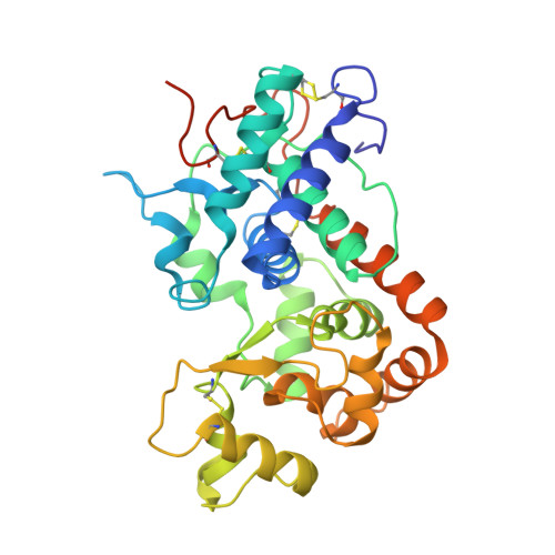
Complexes of Horseradish Peroxidase with Formate, Acetate, and Carbon Monoxide

Carlsson, G.H.Nicholls, P.Svistunenko, D.Berglund, G.I.Hajdu, J.

(2005) Biochemistry 44: 635

  • DOI: 10.1021/bi0483211
  • Primary Citation of Related Structures:  
    1W4W, 1W4Y

  • PubMed Abstract: 
  • Carbon monoxide, formate, and acetate interact with horseradish peroxidase (HRP) by binding to subsites within the active site. These ligands also bind to catalases, but their interactions are different in the two types of enzymes. Formate (notionally the "hydrated" form of carbon monoxide) is oxidized to carbon dioxide by compound I in catalase, while no such reaction is reported to occur in HRP, and the CO complex of ferrocatalase can only be obtained indirectly ...

    Carbon monoxide, formate, and acetate interact with horseradish peroxidase (HRP) by binding to subsites within the active site. These ligands also bind to catalases, but their interactions are different in the two types of enzymes. Formate (notionally the "hydrated" form of carbon monoxide) is oxidized to carbon dioxide by compound I in catalase, while no such reaction is reported to occur in HRP, and the CO complex of ferrocatalase can only be obtained indirectly. Here we describe high-resolution crystal structures for HRP in its complexes with carbon monoxide and with formate, and compare these with the previously determined HRP-acetate structure [Berglund, G. I., et al. (2002) Nature 417, 463-468]. A multicrystal X-ray data collection strategy preserved the correct oxidation state of the iron during the experiments. Absorption spectra of the crystals and electron paramagnetic resonance data for the acetate and formate complexes in solution correlate electronic states with the structural results. Formate in ferric HRP and CO in ferrous HRP bind directly to the heme iron with iron-ligand distances of 2.3 and 1.8 A, respectively. CO does not bind to the ferric iron in the crystal. Acetate bound to ferric HRP stacks parallel with the heme plane with its carboxylate group 3.6 A from the heme iron, and without an intervening solvent molecule between the iron and acetate. The positions of the oxygen atoms in the bound ligands outline a potential access route for hydrogen peroxide to the iron. We propose that interactions in this channel ensure deprotonation of the proximal oxygen before binding to the heme iron.


    Related Citations: 
    • The Catalytic Pathway of Horseradish Peroxidase at High Resolution
      Berglund, G.I., Carlsson, G.H., Smith, A.T., Szoke, H., Henriksen, A., Hajdu, J.
      (2002) Nature 417: 463
    • Expression of a Synthetic Gene for Horseradish Peroxidase C in Escherichia Coli and Folding and Activation of the Recombinant Enzyme with Ca2 and Heme
      Smith, A.T., Santama, N., Dacey, S., Edwards, M., Bray, R.C., Thorneley, R.N., Burke, J.F.
      (1990) J Biol Chem 265: 13335

    Organizational Affiliation

    Molecular Biophysics, Institute of Cellular and Molecular Biology, Uppsala University, Husargatan 3 (Box 596), SE-75124 Uppsala, Sweden.


Macromolecules
Find similar proteins by:  (by identity cutoff)  |  3D Structure
Entity ID: 1
MoleculeChainsSequence LengthOrganismDetailsImage
HORSERADISH PEROXIDASE C1AA323Armoracia rusticanaMutation(s): 0 
Gene Names: PRXC1AHPRC1
EC: 1.11.1.7
UniProt
Find proteins for P00433 (Armoracia rusticana)
Explore P00433 
Go to UniProtKB:  P00433
Entity Groups  
Sequence Clusters30% Identity50% Identity70% Identity90% Identity95% Identity100% Identity
UniProt GroupP00433
Protein Feature View
Expand
  • Reference Sequence
Small Molecules
Ligands 3 Unique
IDChainsName / Formula / InChI Key2D Diagram3D Interactions
HEM
Query on HEM

Download Ideal Coordinates CCD File 
B [auth A]PROTOPORPHYRIN IX CONTAINING FE
C34 H32 Fe N4 O4
KABFMIBPWCXCRK-RGGAHWMASA-L
 Ligand Interaction
FMT
Query on FMT

Download Ideal Coordinates CCD File 
E [auth A]FORMIC ACID
C H2 O2
BDAGIHXWWSANSR-UHFFFAOYSA-N
 Ligand Interaction
CA
Query on CA

Download Ideal Coordinates CCD File 
C [auth A],
D [auth A]
CALCIUM ION
Ca
BHPQYMZQTOCNFJ-UHFFFAOYSA-N
 Ligand Interaction
Experimental Data & Validation

Experimental Data

  • Method: X-RAY DIFFRACTION
  • Resolution: 1.55 Å
  • R-Value Free: 0.215 
  • R-Value Work: 0.198 
  • R-Value Observed: 0.198 
  • Space Group: P 21 21 21
Unit Cell:
Length ( Å )Angle ( ˚ )
a = 40.33α = 90
b = 68.302β = 90
c = 117.048γ = 90
Software Package:
Software NamePurpose
CNSrefinement
DENZOdata reduction
SCALEPACKdata scaling
CNSphasing

Structure Validation

View Full Validation Report


Ligand Structure Quality Assessment 


Entry History 

Deposition Data

Revision History  (Full details and data files)

  • Version 1.0: 2005-01-19
    Type: Initial release
  • Version 1.1: 2011-05-08
    Changes: Version format compliance
  • Version 1.2: 2011-07-13
    Changes: Version format compliance
  • Version 1.3: 2018-01-17
    Changes: Advisory, Data collection
  • Version 1.4: 2019-05-08
    Changes: Data collection, Experimental preparation