Ultra-Fast Barrier-Limited Folding in the Peripheral Subunit-Binding Domain Family.
Ferguson, N., Sharpe, T.D., Schartau, P.J., Sato, S., Allen, M.D., Johnson, C.M., Rutherford, T.J., Fersht, A.R.(2005) J Mol Biol 353: 427
- PubMed: 16168437 Search on PubMed
- DOI: https://doi.org/10.1016/j.jmb.2005.08.031
- Primary Citation Related Structures:
1W4E, 1W4F, 1W4G, 1W4H, 1W4I, 1W4J, 1W4K, 2BTG, 2BTH - PubMed Abstract:
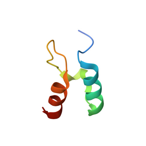
We have determined the solution structures, equilibrium properties and ultra-fast folding kinetics for three bacterial homologues of the peripheral subunit-binding domain (PSBD) family. The mesophilic homologue, BBL, was less stable than the thermophilic and hyper-thermophilic variants (E3BD and POB, respectively). The broad unfolding transitions of each PSBD, when probed by different techniques, were essentially superimposable, consistent with co-operative denaturation. Temperature-jump and continuous-flow fluorescence methods were used to measure the folding kinetics for E3BD, POB and BBL. E3BD folded fairly rapidly at 298K (folding half-time approximately 25 micros) and BBL and POB folded even faster (folding half-times approximately 3-5 micros). The variations in equilibrium and kinetic behaviour observed for the PSBD family resembles that of the homeodomain family, where the folding pattern changes from apparent two-state transitions to multi-state kinetics as the denatured state becomes more structured. The faster folding of POB may be a consequence of its higher propensity to form helical structure in the region corresponding to the folding nucleus of E3BD. The ultra-fast folding of BBL appears to be a consequence of residual structure in the denatured ensemble, as with engrailed homeodomain. We discuss issues concerning "one-state", downhill folding, and find no evidence for, and strong evidence against, it occurring in these PSBDs. The shorter construct used previously for BBL was destabilized significantly and the stability further perturbed by the introduction of fluorescent probes. Thermal titrations for 11 side-chains scattered around the protein, when probed by (13)C-NMR experiments, could be fit globally to a common co-operative transition.
- MRC Centre for Protein Engineering, Hills Road, Cambridge CB2 2QH, UK. nf1@mrc-lmb.cam.ac.uk
Organizational Affiliation: