1W1S

Plant Cytokinin Dehydrogenase in Complex with Benzylaminopurine


Experimental Data Snapshot

  • Method: X-RAY DIFFRACTION
  • Resolution: 2.00 Å
  • R-Value Free: 0.219 
  • R-Value Work: 0.197 
  • R-Value Observed: 0.198 

wwPDB Validation   3D Report Full Report


Ligand Structure Quality Assessment 


This is version 2.0 of the entry. See complete history


Literature

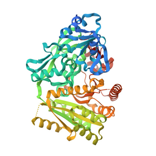
Structures of Michaelis and Product Complexes of Plant Cytokinin Dehydrogenase: Implications for Flavoenzyme Catalysis

Malito, E.Coda, A.Bilyeu, K.Fraaije, M.W.Mattevi, A.

(2004) J Mol Biol 341: 1237

  • DOI: 10.1016/j.jmb.2004.06.083
  • Primary Citation of Related Structures:  
    1W1O, 1W1Q, 1W1R, 1W1S

  • PubMed Abstract: 
  • Cytokinins form a diverse class of compounds that are essential for plant growth. Cytokinin dehydrogenase has a major role in the control of the levels of these plant hormones by catalysing their irreversible oxidation. The crystal structure of Zea mays cytokinin dehydrogenase displays the same two-domain topology of the flavoenzymes of the vanillyl-alcohol oxidase family but its active site cannot be related to that of any other family member ...

    Cytokinins form a diverse class of compounds that are essential for plant growth. Cytokinin dehydrogenase has a major role in the control of the levels of these plant hormones by catalysing their irreversible oxidation. The crystal structure of Zea mays cytokinin dehydrogenase displays the same two-domain topology of the flavoenzymes of the vanillyl-alcohol oxidase family but its active site cannot be related to that of any other family member. The X-ray analysis reveals a bipartite architecture of the catalytic centre, which consists of a funnel-shaped region on the protein surface and an internal cavity lined by the flavin ring. A pore with diameter of about 4A connects the two active-site regions. Snapshots of two critical steps along the reaction cycle were obtained through the structural analysis of the complexes with a slowly reacting substrate and the reaction product, which correspond to the states immediately before (Michaelis complex) and after (product complex) oxidation has taken place. The substrate displays a "plug-into-socket" binding mode that seals the catalytic site and precisely positions the carbon atom undergoing oxidation in close contact with the reactive locus of the flavin. A polarising H-bond between the substrate amine group and an Asp-Glu pair may facilitate oxidation. Substrate to product conversion results in small atomic movements, which lead to a planar conformation of the reaction product allowing double-bond conjugation. These features in the mechanism of amine recognition and oxidation differ from those observed in other flavin-dependent amine oxidases.


    Organizational Affiliation

    Department of Genetics and Microbiology, University of Pavia, Via Ferrata 1, 27100 Pavia, Italy.


Macromolecules
Find similar proteins by:  (by identity cutoff)  |  3D Structure
Entity ID: 1
MoleculeChainsSequence LengthOrganismDetailsImage
CYTOKININ DEHYDROGENASEA534Zea maysMutation(s): 0 
Gene Names: CKX1
EC: 1.5.99.12
UniProt
Find proteins for Q9T0N8 (Zea mays)
Explore Q9T0N8 
Go to UniProtKB:  Q9T0N8
Entity Groups  
Sequence Clusters30% Identity50% Identity70% Identity90% Identity95% Identity100% Identity
UniProt GroupQ9T0N8
Protein Feature View
Expand
  • Reference Sequence
Oligosaccharides

Help

Entity ID: 2
MoleculeChainsChain Length2D DiagramGlycosylation3D Interactions
2-acetamido-2-deoxy-beta-D-glucopyranose-(1-4)-2-acetamido-2-deoxy-beta-D-glucopyranoseB 2N-Glycosylation Oligosaccharides Interaction
Glycosylation Resources
GlyTouCan:  G42666HT
GlyCosmos:  G42666HT
GlyGen:  G42666HT
Small Molecules
Ligands 3 Unique
IDChainsName / Formula / InChI Key2D Diagram3D Interactions
FAD
Query on FAD

Download Ideal Coordinates CCD File 
C [auth A]FLAVIN-ADENINE DINUCLEOTIDE
C27 H33 N9 O15 P2
VWWQXMAJTJZDQX-UYBVJOGSSA-N
 Ligand Interaction
EMU
Query on EMU

Download Ideal Coordinates CCD File 
D [auth A]N-BENZYL-9H-PURIN-6-AMINE
C12 H11 N5
NWBJYWHLCVSVIJ-UHFFFAOYSA-N
 Ligand Interaction
NAG
Query on NAG

Download Ideal Coordinates CCD File 
E [auth A],
F [auth A],
G [auth A]
2-acetamido-2-deoxy-beta-D-glucopyranose
C8 H15 N O6
OVRNDRQMDRJTHS-FMDGEEDCSA-N
 Ligand Interaction
Experimental Data & Validation

Experimental Data

  • Method: X-RAY DIFFRACTION
  • Resolution: 2.00 Å
  • R-Value Free: 0.219 
  • R-Value Work: 0.197 
  • R-Value Observed: 0.198 
  • Space Group: P 42 21 2
Unit Cell:
Length ( Å )Angle ( ˚ )
a = 168.683α = 90
b = 168.683β = 90
c = 53.759γ = 90
Software Package:
Software NamePurpose
REFMACrefinement
MOSFLMdata reduction
SCALAdata scaling
CCP4phasing

Structure Validation

View Full Validation Report


Ligand Structure Quality Assessment 


Entry History 

Deposition Data

Revision History  (Full details and data files)

  • Version 1.0: 2004-08-26
    Type: Initial release
  • Version 1.1: 2011-05-08
    Changes: Version format compliance
  • Version 1.2: 2011-07-13
    Changes: Version format compliance
  • Version 2.0: 2020-07-29
    Type: Remediation
    Reason: Carbohydrate remediation
    Changes: Atomic model, Data collection, Derived calculations, Other, Structure summary