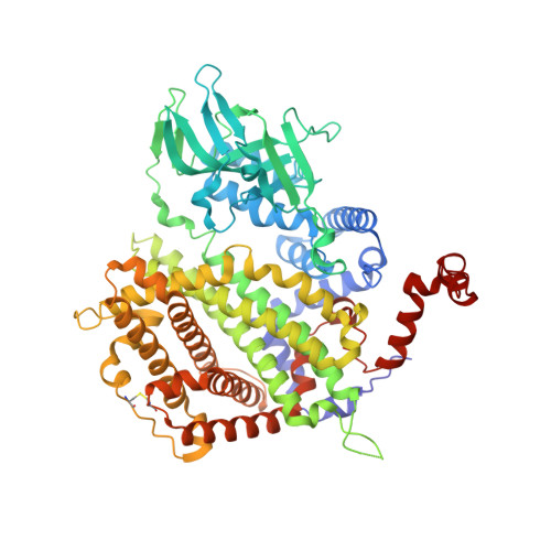
1W07 | pdb_00001w07

Arabidopsis thaliana acyl-CoA oxidase 1


Experimental Data Snapshot

  • Method: X-RAY DIFFRACTION
  • Resolution: 2.00 Å
  • R-Value Free: 
    0.249 (Depositor), 0.240 (DCC) 
  • R-Value Work: 
    0.204 (Depositor), 0.200 (DCC) 
  • R-Value Observed: 
    0.204 (Depositor) 

Starting Model: experimental
View more details

wwPDB Validation   3D Report Full Report


Ligand Structure Quality Assessment 

Created with Raphaël 2.3.0Worse 01 BetterLigand structure goodness of fit to experimental dataBest fitted FADClick on this verticalbar to view details

This is version 1.5 of the entry. See complete history


Literature

Acyl-Coa Oxidase 1 from Arabidopsis Thaliana. Structure of a Key Enzyme in Plant Lipid Metabolism

Pedersen, L.Henriksen, A.

(2005) J Mol Biology 345: 487

  • DOI: https://doi.org/10.1016/j.jmb.2004.10.062
  • Primary Citation of Related Structures:  
    1W07

  • PubMed Abstract: 

    The peroxisomal acyl-CoA oxidase family plays an essential role in lipid metabolism by catalyzing the conversion of acyl-CoA into trans-2-enoyl-CoA during fatty acid beta-oxidation. Here, we report the X-ray structure of the FAD-containing Arabidopsis thaliana acyl-CoA oxidase 1 (ACX1), the first three-dimensional structure of a plant acyl-CoA oxidase. Like other acyl-CoA oxidases, the enzyme is a dimer and it has a fold resembling that of mammalian acyl-CoA oxidase. A comparative analysis including mammalian acyl-CoA oxidase and the related tetrameric mitochondrial acyl-CoA dehydrogenases reveals a substrate-binding architecture that explains the observed preference for long-chained, mono-unsaturated substrates in ACX1. Two anions are found at the ACX1 dimer interface and for the first time the presence of a disulfide bridge in a peroxisomal protein has been observed. The functional differences between the peroxisomal acyl-CoA oxidases and the mitochondrial acyl-CoA dehydrogenases are attributed to structural differences in the FAD environments.


  • Organizational Affiliation

    Carlsberg Laboratory, Gamle Carlsberg Vej 10, DK-2500 Valby, Denmark.


Macromolecules
Find similar proteins by:  (by identity cutoff)  |  3D Structure
Entity ID: 1
MoleculeChains Sequence LengthOrganismDetailsImage
ACYL-COA OXIDASE
A, B
659Arabidopsis thalianaMutation(s): 1 
EC: 1.3.3.6
UniProt
Find proteins for O65202 (Arabidopsis thaliana)
Explore O65202 
Go to UniProtKB:  O65202
Entity Groups  
Sequence Clusters30% Identity50% Identity70% Identity90% Identity95% Identity100% Identity
UniProt GroupO65202
Sequence Annotations
Expand
  • Reference Sequence
Small Molecules
Ligands 4 Unique
IDChains Name / Formula / InChI Key2D Diagram3D Interactions
FAD
Query on FAD

Download Ideal Coordinates CCD File 
C [auth A],
I [auth B]
FLAVIN-ADENINE DINUCLEOTIDE
C27 H33 N9 O15 P2
VWWQXMAJTJZDQX-UYBVJOGSSA-N
PT
Query on PT

Download Ideal Coordinates CCD File 
F [auth A]
G [auth A]
H [auth A]
K [auth B]
L [auth B]
PLATINUM (II) ION
Pt
HRGDZIGMBDGFTC-UHFFFAOYSA-N
CA
Query on CA

Download Ideal Coordinates CCD File 
D [auth A]CALCIUM ION
Ca
BHPQYMZQTOCNFJ-UHFFFAOYSA-N
CL
Query on CL

Download Ideal Coordinates CCD File 
E [auth A],
J [auth B]
CHLORIDE ION
Cl
VEXZGXHMUGYJMC-UHFFFAOYSA-M
Experimental Data & Validation

Experimental Data

  • Method: X-RAY DIFFRACTION
  • Resolution: 2.00 Å
  • R-Value Free:  0.249 (Depositor), 0.240 (DCC) 
  • R-Value Work:  0.204 (Depositor), 0.200 (DCC) 
  • R-Value Observed: 0.204 (Depositor) 
Space Group: P 21 21 21
Unit Cell:
Length ( Å )Angle ( ˚ )
a = 85.201α = 90
b = 117.001β = 90
c = 131.047γ = 90
Software Package:
Software NamePurpose
CNSrefinement
MOSFLMdata reduction
SCALAdata scaling

Structure Validation

View Full Validation Report


Ligand Structure Quality Assessment 

Created with Raphaël 2.3.0Worse 01 BetterLigand structure goodness of fit to experimental dataBest fitted FADClick on this verticalbar to view details

Entry History 

Deposition Data

Revision History  (Full details and data files)

  • Version 1.0: 2004-12-15
    Type: Initial release
  • Version 1.1: 2011-05-08
    Changes: Version format compliance
  • Version 1.2: 2011-07-13
    Changes: Version format compliance
  • Version 1.3: 2018-01-17
    Changes: Data collection
  • Version 1.4: 2023-12-13
    Changes: Data collection, Database references, Derived calculations, Other, Refinement description
  • Version 1.5: 2024-11-06
    Changes: Structure summary