1VZU | pdb_00001vzu

Roles of active site tryptophans in substrate binding and catalysis by ALPHA-1,3 GALACTOSYLTRANSFERASE


Experimental Data Snapshot

  • Method: X-RAY DIFFRACTION
  • Resolution: 1.97 Å
  • R-Value Free: 
    0.202 (Depositor) 
  • R-Value Work: 
    0.154 (Depositor) 
  • R-Value Observed: 
    0.154 (Depositor) 

Starting Model: experimental
View more details

wwPDB Validation 3D Report Full Report

Validation slider image for 1VZU

This is version 2.1 of the entry. See complete history

Literature

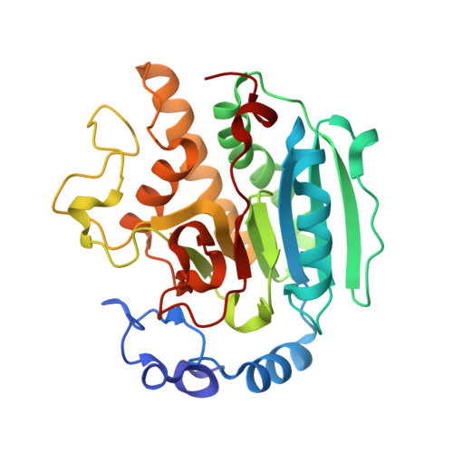
Roles of active site tryptophans in substrate binding and catalysis by alpha-1,3 galactosyltransferase.

Zhang, Y.Deshpande, A.Xie, Z.Natesh, R.Acharya, K.R.Brew, K.

(2004) Glycobiology 14: 1295-1302

  • DOI: https://doi.org/10.1093/glycob/cwh119
  • Primary Citation Related Structures: 
    1VZU, 1VZX

  • PubMed Abstract: 

    Aromatic amino acids are frequent components of the carbohydrate binding sites of lectins and enzymes. Previous structural studies have shown that in alpha-1,3 galactosyltransferase, the binding site for disaccharide acceptor substrates is encircled by four tryptophans, residues 249, 250, 314, and 356. To investigate their roles in enzyme specificity and catalysis, we expressed and characterized variants of the catalytic domain of alpha-1,3 galactosyltransferase with substitutions for each tryptophan. Substitution of glycine for tryptophan 249, whose indole ring interacts with the nonpolar B face of glucose or GlcNAc, greatly increases the K(m) for the acceptor substrate. In contrast, the substitution of tyrosine for tryptophan 314, which interacts with the beta-galactosyl moiety of the acceptor and UDP-galactose, decreases k(cat) for the galactosyltransferase reaction but does not affect the low UDP-galactose hydrolase activity. Thus, this highly conserved residue stabilizes the transition state for the galactose transfer to disaccharide but not to water. High-resolution crystallographic structures of the Trp(249)Gly mutant and the Trp(314)Tyr mutant indicate that the mutations do not affect the overall structure of the enzyme or its interactions with ligands. Substitutions for tryptophan 250 have only small effects on catalytic activity, but mutation of tryptophan 356 to threonine reduces catalytic activity for both transferase and hydrolase activities and reduces affinity for the acceptor substrate. This residue is adjacent to the flexible C-terminus that becomes ordered on binding UDP to assemble the acceptor binding site and influence catalysis. The results highlight the diverse roles of these tryptophans in enzyme action and the importance of k(cat) changes in modulating glycosyltransferase specificity.


  • Organizational Affiliation
    • Department of Biomedical Sciences, Florida Atlantic University, Boca Raton, FL 33341, USA.

Macromolecule Content 

  • Total Structure Weight: 69.92 kDa 
  • Atom Count: 5,788 
  • Modeled Residue Count: 574 
  • Deposited Residue Count: 578 
  • Unique protein chains: 1

Macromolecules

Find similar proteins by:|  3D Structure
Entity ID: 1
MoleculeChains  Sequence LengthOrganismDetailsImage
N-ACETYLLACTOSAMINIDE ALPHA-1,3-GALACTOSYLTRANSFERASE
A, B
289Bos taurusMutation(s): 1 
EC: 2.4.1.151 (PDB Primary Data), 2.4.1.87 (UniProt)
UniProt
Find proteins for P14769 (Bos taurus)
Explore P14769 
Go to UniProtKB:  P14769
Entity Groups
Sequence Clusters30% Identity50% Identity70% Identity90% Identity95% Identity100% Identity
UniProt GroupP14769
Sequence Annotations
Expand
Reference Sequence

Oligosaccharides

Help  
Entity ID: 2
MoleculeChains Length2D Diagram GlycosylationD Interactions
beta-D-galactopyranose-(1-4)-beta-D-glucopyranose
C, D
2N/A
Glycosylation Resources
GlyTouCan: G84224TW
GlyCosmos: G84224TW
GlyGen: G84224TW

Biologically Interesting Molecules (External Reference) 

1 Unique

Experimental Data & Validation

Experimental Data

  • Method: X-RAY DIFFRACTION
  • Resolution: 1.97 Å
  • R-Value Free:  0.202 (Depositor) 
  • R-Value Work:  0.154 (Depositor) 
  • R-Value Observed: 0.154 (Depositor) 
Space Group: P 1 21 1
Unit Cell:
Length ( Å )Angle ( ˚ )
a = 44.858α = 90
b = 93.684β = 99
c = 93.714γ = 90
Software Package:
Software NamePurpose
CNSrefinement
DENZOdata reduction
SCALEPACKdata scaling
AMoREphasing

Structure Validation

View Full Validation Report


Entry History 

Deposition Data

Revision History  (Full details and data files)

  • Version 1.0: 2004-07-08
    Type: Initial release
  • Version 1.1: 2011-05-08
    Changes: Version format compliance
  • Version 1.2: 2011-07-13
    Changes: Version format compliance
  • Version 1.3: 2018-02-28
    Changes: Database references, Source and taxonomy
  • Version 2.0: 2020-07-29
    Type: Remediation
    Reason: Carbohydrate remediation
    Changes: Atomic model, Data collection, Derived calculations, Non-polymer description, Structure summary
  • Version 2.1: 2023-12-13
    Changes: Data collection, Database references, Refinement description, Structure summary