The three-dimensional solution structure of the NK-2 homeodomain from Drosophila.
Tsao, D.H., Gruschus, J.M., Wang, L.H., Nirenberg, M., Ferretti, J.A.(1995) J Mol Biol 251: 297-307
- PubMed: 7643404 Search on PubMed
- DOI: https://doi.org/10.1006/jmbi.1995.0435
- Primary Citation Related Structures:
1VND - PubMed Abstract:
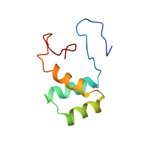
We describe the NMR determination of the three-dimensional structure of a 77 amino acid residue protein, which consists of the 60 residue NK-2 homeodomain from Drosophila melanogaster and adjacent amino acid residues. The NK-2 homeodomain protein is part of a 723 amino acid residue protein which is expressed early in embryonic development in part of the central nervous system. NK-2 was characterized using both a natural abundance and a uniformly 15N enriched sample by two-dimensional and three-dimensional NMR experiments. The average root-mean-square deviation for 30 structures for residues 8 to 53 is 0.40 A for the backbone heavy-atoms and 0.72 A for the backbone and side-chain heavy-atoms. These structures were obtained from 986 NOE-derived upper and lower bound restraints. The three-dimensional structure contains three helices which consist of homeodomain amino acid residues 10 to 22, 28 to 38 and 42 to 52, as well as a turn between helix II and III, characteristic of homeodomains. Residues 53 to 60 of the DNA recognition helix are not fully ordered in the absence of DNA. In the free state this segment adopts a flexible but helix-like structure between residues 53 and 56 and is disordered from residues 57 to 60 although, as shown previously, the helix elongates by eight residues upon binding to DNA. The role of variable residues 52, 54 and 56 in determining the structure and flexibility of the recognition helix, as well as the stability of the NK-2 homeodomain as manifested by its thermal denaturation, are discussed.
- Laboratory of Biophysical Chemistry, National Heart, Lung and Blood Institute, National Institutes of Health, Bethesda, MD 20892, USA.
Organizational Affiliation: