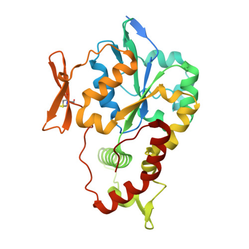
Crystal structure and mutational analysis of heparan sulfate 3-O-sulfotransferase isoform 1
Edavettal, S.C., Lee, K.A., Negishi, M., Linhardt, R.J., Liu, J., Pedersen, L.C.(2004) J Biol Chem 279: 25789-25797
- PubMed: 15060080 Search on PubMed
- DOI: https://doi.org/10.1074/jbc.M401089200
- Primary Citation Related Structures:
1VKJ - PubMed Abstract:
Heparan sulfate interacts with antithrombin, a protease inhibitor, to regulate blood coagulation. Heparan sulfate 3-O-sulfotransferase isoform 1 performs the crucial last step modification in the biosynthesis of anticoagulant heparan sulfate. This enzyme transfers the sulfuryl group (SO(3)) from 3'-phosphoadenosine 5'-phosphosulfate to the 3-OH position of a glucosamine residue to form the 3-O-sulfo glucosamine, a structural motif critical for binding of heparan sulfate to antithrombin. In this study, we report the crystal structure of 3-O-sulfotransferase isoform 1 at 2.5-A resolution in a binary complex with 3'-phosphoadenosine 5'-phosphate. This structure reveals residues critical for 3'-phosphoadenosine 5'-phosphosulfate binding and suggests residues required for the binding of heparan sulfate. In addition, site-directed mutagenesis analyses suggest that residues Arg-67, Lys-68, Arg-72, Glu-90, His-92, Asp-95, Lys-123, and Arg-276 are essential for enzymatic activity. Among these essential amino acid residues, we find that residues Arg-67, Arg-72, His-92, and Asp-95 are conserved in heparan sulfate 3-O-sulfotransferases but not in heparan N-deacetylase/N-sulfotransferase, suggesting a role for these residues in conferring substrate specificity. Results from this study provide information essential for understanding the biosynthesis of anticoagulant heparan sulfate and the general mechanism of action of heparan sulfate sulfotransferases.
- Division of Medicinal Chemistry and Natural Products, School of Pharmacy, University of North Carolina, Chapel Hill, North Carolina 27599, USA.
Organizational Affiliation: