1VF6

2.1 Angstrom crystal structure of the PALS-1-L27N and PATJ L27 heterodimer complex


Experimental Data Snapshot

  • Method: X-RAY DIFFRACTION
  • Resolution: 2.10 Å
  • R-Value Free: 0.262 
  • R-Value Work: 0.242 
  • R-Value Observed: 0.242 

wwPDB Validation   3D Report Full Report


This is version 1.4 of the entry. See complete history


Literature

Structural basis for L27 domain-mediated assembly of signaling and cell polarity complexes.

Li, Y.Karnak, D.Demeler, B.Margolis, B.Lavie, A.

(2004) EMBO J 23: 2723-2733

  • DOI: https://doi.org/10.1038/sj.emboj.7600294
  • Primary Citation of Related Structures:  
    1VF6

  • PubMed Abstract: 

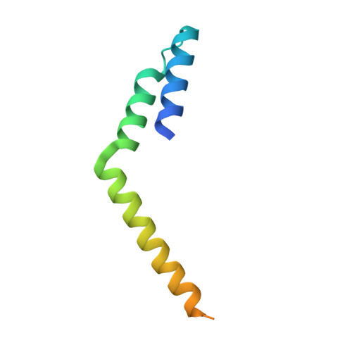
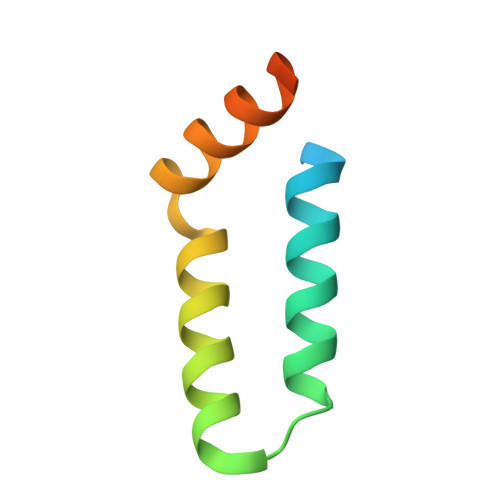
    L27 is a protein-binding domain that can assemble essential proteins for signaling and cell polarity into complexes by interacting in a heterodimeric manner. One of these protein complexes is the PATJ/PALS1/Crumbs tripartite complex, which is crucial for the establishment and maintenance of cell polarity. To reveal the structural basis underlining the obligate heterodimerization, we have determined the crystal structure of the PALS1-L27N/PATJ-L27 heterodimer complex. Each L27 domain is composed of three helices. The two L27 domains heterodimerize by building a compact structure consisting of a four-helix bundle formed by the first two helices of each L27 domain and one coiled-coil formed by the third helix of each domain. The large hydrophobic packing interactions contributed by all the helices of both L27 domains predominantly drive the heterodimer formation, which is likely to be a general feature of L27 domains. Combined with mutational studies, we can begin to understand the structural basis for the specificity of L27 binding pairs. Our results provide unique insights into L27 domain heterodimer complex, which is critical for cell polarization.


  • Organizational Affiliation

    Department of Biochemistry and Molecular Genetics, University of Illinois, Chicago, IL 60607, USA.


Macromolecules
Find similar proteins by:  (by identity cutoff)  |  3D Structure
Entity ID: 1
MoleculeChains Sequence LengthOrganismDetailsImage
PALS1-associated tight junction protein
A, B
83Homo sapiensMutation(s): 0 
Membrane Entity: Yes 
UniProt & NIH Common Fund Data Resources
Find proteins for Q8NI35 (Homo sapiens)
Explore Q8NI35 
Go to UniProtKB:  Q8NI35
PHAROS:  Q8NI35
GTEx:  ENSG00000132849 
Entity Groups  
Sequence Clusters30% Identity50% Identity70% Identity90% Identity95% Identity100% Identity
UniProt GroupQ8NI35
Sequence Annotations
Expand
  • Reference Sequence
Find similar proteins by:  (by identity cutoff)  |  3D Structure
Entity ID: 2
MoleculeChains Sequence LengthOrganismDetailsImage
MAGUK p55 subfamily member 5
C, D
72Mus musculusMutation(s): 0 
Membrane Entity: Yes 
UniProt
Find proteins for Q9JLB2 (Mus musculus)
Explore Q9JLB2 
Go to UniProtKB:  Q9JLB2
Entity Groups  
Sequence Clusters30% Identity50% Identity70% Identity90% Identity95% Identity100% Identity
UniProt GroupQ9JLB2
Sequence Annotations
Expand
  • Reference Sequence
Experimental Data & Validation

Experimental Data

  • Method: X-RAY DIFFRACTION
  • Resolution: 2.10 Å
  • R-Value Free: 0.262 
  • R-Value Work: 0.242 
  • R-Value Observed: 0.242 
  • Space Group: H 3 2
Unit Cell:
Length ( Å )Angle ( ˚ )
a = 111.06α = 90
b = 111.06β = 90
c = 193.69γ = 120
Software Package:
Software NamePurpose
HKL-2000data collection
XDSdata reduction
SOLVEphasing
CNSrefinement
HKL-2000data reduction
XDSdata scaling

Structure Validation

View Full Validation Report


Entry History 

Deposition Data

Revision History  (Full details and data files)

  • Version 1.0: 2004-04-20
    Type: Initial release
  • Version 1.1: 2008-04-27
    Changes: Version format compliance
  • Version 1.2: 2011-07-13
    Changes: Version format compliance
  • Version 1.3: 2016-12-07
    Changes: Other
  • Version 1.4: 2023-12-27
    Changes: Data collection, Database references