


Crystal structure of anthranilate phosphoribosyltransferase (TrpD) from Thermus thermophilus HB8
Shimizu, K., Kunishima, N.To be published.
Experimental Data Snapshot
Starting Model: experimental
View more details
wwPDB Validation 3D Report Full Report

Entity ID: 1 | |||||
|---|---|---|---|---|---|
| Molecule | Chains | Sequence Length | Organism | Details | Image |
| anthranilate phosphoribosyltransferase | 329 | Thermus thermophilus | Mutation(s): 0 EC: 2.4.2.18 | 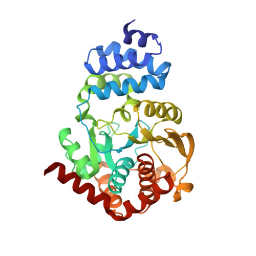 | |
UniProt | |||||
Entity Groups | |||||
| Sequence Clusters | 30% Identity50% Identity70% Identity90% Identity95% Identity100% Identity | ||||
| UniProt Group | P83827 | ||||
Sequence AnnotationsExpand | |||||
Reference Sequence | |||||
| Length ( Å ) | Angle ( ˚ ) |
|---|---|
| a = 143.318 | α = 90 |
| b = 72.45 | β = 110.36 |
| c = 73.539 | γ = 90 |
| Software Name | Purpose |
|---|---|
| CNS | refinement |
| HKL-2000 | data reduction |
| SCALEPACK | data scaling |
| AMoRE | phasing |


