Structural basis of human cytoglobin for ligand binding.
Sugimoto, H., Makino, M., Sawai, H., Kawada, N., Yoshizato, K., Shiro, Y.(2004) J Mol Biol 339: 873-885
- PubMed: 15165856 Search on PubMed
- DOI: https://doi.org/10.1016/j.jmb.2004.04.024
- Primary Citation Related Structures:
1V5H - PubMed Abstract:
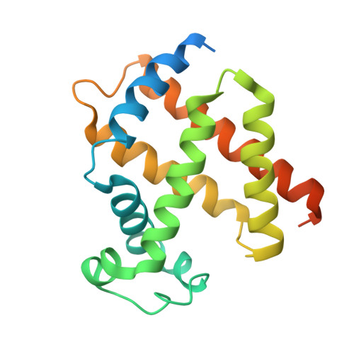
Cytoglobin (Cgb), a newly discovered member of the vertebrate globin family, binds O(2) reversibly via its heme, as is the case for other mammalian globins (hemoglobin (Hb), myoglobin (Mb) and neuroglobin (Ngb)). While Cgb is expressed in various tissues, its physiological role is not clearly understood. Here, the X-ray crystal structure of wild type human Cgb in the ferric state at 2.4A resolution is reported. In the crystal structure, ferric Cgb is dimerized through two intermolecular disulfide bonds between Cys38(B2) and Cys83(E9), and the dimerization interface is similar to that of lamprey Hb and Ngb. The overall backbone structure of the Cgb monomer exhibits a traditional globin fold with a three-over-three alpha-helical sandwich, in which the arrangement of helices is basically the same among all globins studied to date. A detailed comparison reveals that the backbone structure of the CD corner to D helix region, the N terminus of the E-helix and the F-helix of Cgb resembles more closely those of pentacoordinated globins (Mb, lamprey Hb), rather than hexacoordinated globins (Ngb, rice Hb). However, the His81(E7) imidazole group coordinates directly to the heme iron as a sixth axial ligand to form a hexcoordinated heme, like Ngb and rice Hb. The position and orientation of the highly conserved residues in the heme pocket (Phe(CD1), Val(E11), distal His(E7) and proximal His(F8)) are similar to those of other globin proteins. Two alternative conformations of the Arg84(E10) guanidium group were observed, suggesting that it participates in ligand binding to Cgb, as is the case for Arg(E10) of Aplysia Mb and Lys(E10) of Ngb. The structural diversities and similarities among globin proteins are discussed with relevance to molecular evolutionary relationships.
- Biophysical Chemistry Laboratory, Riken Harima Institute/SPring8, Hyogo 679-5148, Japan.
Organizational Affiliation: