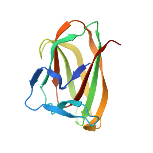
The Crystal Structure of the Family 6 Carbohydrate Binding Module from Cellvibrio Mixtus Endoglucanase 5A in Complex with Oligosaccharides Reveals Two Distinct Binding Sites with Different Ligand Specificities
Pires, V.M.R., Henshaw, J., Prates, J.A.M., Bolam, D., Ferreira, L.M.A., Fontes, C.M.G.A., Henrissat, B., Planas, A., Gilbert, H.J., Czjzek, M.(2004) J Biological Chem 279: 21560
- PubMed: 15010454
- DOI: https://doi.org/10.1074/jbc.M401599200
- Primary Citation of Related Structures:
1UXX, 1UXZ, 1UY0, 1UYX, 1UYY, 1UYZ, 1UZ0 - PubMed Abstract:
Glycoside hydrolases that release fixed carbon from the plant cell wall are of considerable biological and industrial importance. These hydrolases contain non-catalytic carbohydrate binding modules (CBMs) that, by bringing the appended catalytic domain into intimate association with its insoluble substrate, greatly potentiate catalysis. Family 6 CBMs (CBM6) are highly unusual because they contain two distinct clefts (cleft A and cleft B) that potentially can function as binding sites. Henshaw et al. (Henshaw, J., Bolam, D. N., Pires, V. M. R., Czjzek, M., Henrissat, B., Ferreira, L. M. A., Fontes, C. M. G. A., and Gilbert, H. J. (2003) J. Biol. Chem. 279, 21552-21559) show that CmCBM6 contains two binding sites that display both similarities and differences in their ligand specificity. Here we report the crystal structure of CmCBM6 in complex with a variety of ligands that reveals the structural basis for the ligand specificity displayed by this protein. In cleft A the two faces of the terminal sugars of beta-linked oligosaccharides stack against Trp-92 and Tyr-33, whereas the rest of the binding cleft is blocked by Glu-20 and Thr-23, residues that are not present in CBM6 proteins that bind to the internal regions of polysaccharides in cleft A. Cleft B is solvent-exposed and, therefore, able to bind ligands because the loop, which occludes this region in other CBM6 proteins, is much shorter and flexible (lacks a conserved proline) in CmCBM6. Subsites 2 and 3 of cleft B accommodate cellobiose (Glc-beta-1,4-Glc), subsite 4 will bind only to a beta-1,3-linked glucose, whereas subsite 1 can interact with either a beta-1,3- or beta-1,4-linked glucose. These different specificities of the subsites explain how cleft B can accommodate beta-1,4-beta-1,3- or beta-1,3-beta-1,4-linked gluco-configured ligands.
- CIISA-Faculadade de Medicina Veterinaria, Rua Prof. Cid dos Santos, 1300-477 Lisbon, Portugal.
Organizational Affiliation:


