1UW4

The structural basis of the interaction between nonsense mediated decay factors UPF2 and UPF3


Experimental Data Snapshot

  • Method: X-RAY DIFFRACTION
  • Resolution: 1.95 Å
  • R-Value Free: 0.227 
  • R-Value Work: 0.204 
  • R-Value Observed: 0.205 

wwPDB Validation   3D Report Full Report


This is version 1.1 of the entry. See complete history


Literature

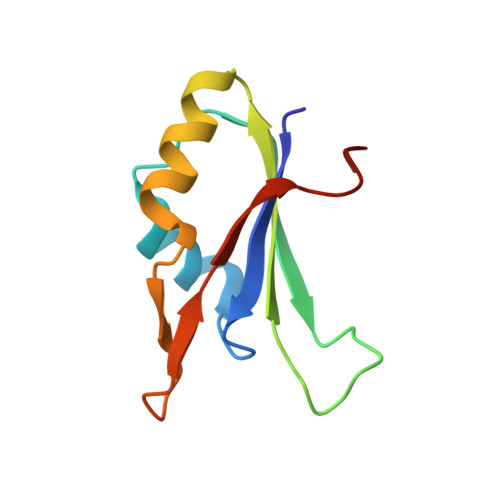
The Structural Basis for the Interaction between Nonsense-Mediated Mrna Decay Factors Upf2 and Upf3

Kadlec, J.Izaurralde, E.Cusack, S.

(2004) Nat Struct Mol Biol 11: 330

  • DOI: https://doi.org/10.1038/nsmb741
  • Primary Citation of Related Structures:  
    1UW4

  • PubMed Abstract: 

    Nonsense-mediated mRNA decay (NMD) is a surveillance mechanism by which eukaryotic cells detect and degrade transcripts containing premature termination codons. Three 'up-frameshift' proteins, UPF1, UPF2 and UPF3, are essential for this process in organisms ranging from yeast to human. We present a crystal structure at a resolution of 1.95 A of the complex between the interacting domains of human UPF2 and UPF3b, which are, respectively, a MIF4G (middle portion of eIF4G) domain and an RNP domain (ribonucleoprotein-type RNA-binding domain). The protein-protein interface is mediated by highly conserved charged residues in UPF2 and UPF3b and involves the beta-sheet surface of the UPF3b RNP domain, which is generally used by these domains to bind nucleic acids. We show that the UPF3b RNP does not bind RNA, whereas the UPF2 construct and the complex do. Our results advance understanding of the molecular mechanisms underlying the NMD quality control process.


  • Organizational Affiliation

    European Molecular Biology Laboratory, Grenoble Outstation, 6 rue Jules Horowitz, BP 181, F-38042 Grenoble Cedex 9, France.


Macromolecules
Find similar proteins by:  (by identity cutoff)  |  3D Structure
Entity ID: 1
MoleculeChains Sequence LengthOrganismDetailsImage
UPF3X
A, C
91Homo sapiensMutation(s): 0 
UniProt & NIH Common Fund Data Resources
Find proteins for Q9BZI7 (Homo sapiens)
Explore Q9BZI7 
Go to UniProtKB:  Q9BZI7
PHAROS:  Q9BZI7
GTEx:  ENSG00000125351 
Entity Groups  
Sequence Clusters30% Identity50% Identity70% Identity90% Identity95% Identity100% Identity
UniProt GroupQ9BZI7
Sequence Annotations
Expand
  • Reference Sequence
Find similar proteins by:  (by identity cutoff)  |  3D Structure
Entity ID: 2
MoleculeChains Sequence LengthOrganismDetailsImage
REGULATOR OF NONSENSE TRANSCRIPTS 2
B, D
248Homo sapiensMutation(s): 0 
UniProt & NIH Common Fund Data Resources
Find proteins for Q9HAU5 (Homo sapiens)
Explore Q9HAU5 
Go to UniProtKB:  Q9HAU5
GTEx:  ENSG00000151461 
Entity Groups  
Sequence Clusters30% Identity50% Identity70% Identity90% Identity95% Identity100% Identity
UniProt GroupQ9HAU5
Sequence Annotations
Expand
  • Reference Sequence
Small Molecules
Experimental Data & Validation

Experimental Data

  • Method: X-RAY DIFFRACTION
  • Resolution: 1.95 Å
  • R-Value Free: 0.227 
  • R-Value Work: 0.204 
  • R-Value Observed: 0.205 
  • Space Group: P 21 21 2
Unit Cell:
Length ( Å )Angle ( ˚ )
a = 70.306α = 90
b = 100.178β = 90
c = 153.458γ = 90
Software Package:
Software NamePurpose
REFMACrefinement
XDSdata reduction
XSCALEdata scaling
SOLVE/RESOLVEphasing

Structure Validation

View Full Validation Report


Entry History 

Deposition Data

Revision History  (Full details and data files)

  • Version 1.0: 2004-03-11
    Type: Initial release
  • Version 1.1: 2011-07-13
    Changes: Advisory, Version format compliance