Bar Domains as Sensors of Membrane Curvature: The Amphiphysin Bar Structure
Peter, B.J., Kent, H.M., Mills, I.G., Vallis, Y., Butler, J.G., Evans, P.R., Mcmahon, H.T.(2004) Science 303: 495
- PubMed: 14645856 Search on PubMed
- DOI: https://doi.org/10.1126/science.1092586
- Primary Citation Related Structures:
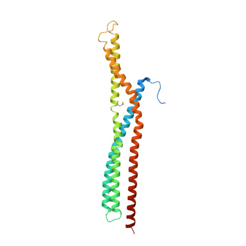
1URU - PubMed Abstract:
The BAR (Bin/amphiphysin/Rvs) domain is the most conserved feature in amphiphysins from yeast to human and is also found in endophilins and nadrins. We solved the structure of the Drosophila amphiphysin BAR domain. It is a crescent-shaped dimer that binds preferentially to highly curved negatively charged membranes. With its N-terminal amphipathic helix and BAR domain (N-BAR), amphiphysin can drive membrane curvature in vitro and in vivo. The structure is similar to that of arfaptin2, which we find also binds and tubulates membranes. From this, we predict that BAR domains are in many protein families, including sorting nexins, centaurins, and oligophrenins. The universal and minimal BAR domain is a dimerization, membrane-binding, and curvature-sensing module.
- Medical Research Council (MRC) Laboratory of Molecular Biology, Hills Road, Cambridge CB2 2QH, UK.
Organizational Affiliation: