Nad+ and Metal-Ion Dependent Hydrolysis by Family 4 Glycosidases: Structural Insight Into Specificity for Phospho-Beta-D-Glucosides
Varrot, A., Yip, V.L., Li, Y., Rajan, S.S., Yang, X., Anderson, W.F., Thompson, J., Withers, S.G., Davies, G.J.(2005) J Mol Biology 346: 423
- PubMed: 15670594 Search on PubMed
- DOI: https://doi.org/10.1016/j.jmb.2004.11.058
- Primary Citation Related Structures:
1UP4, 1UP7 - PubMed Abstract:
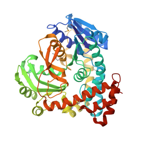
The import of disaccharides by many bacteria is achieved through their simultaneous translocation and phosphorylation by the phosphoenolpyruvate-dependent phosphotransferase system (PEP-PTS). The imported phospho-disaccharides are, in some cases, subsequently hydrolyzed by members of the unusual glycoside hydrolase family GH4. The GH4 enzymes, occasionally found also in bacteria such as Thermotoga maritima that do not utilise a PEP-PTS system, require both NAD(+) and Mn(2+) for catalysis. A further curiosity of this family is that closely related enzymes may show specificity for either alpha-d- or beta-d-glycosides. Here, we present, for the first time, the three-dimensional structure (using single-wavelength anomalous dispersion methods, harnessing extensive non-crystallographic symmetry) of the 6-phospho-beta-glycosidase, BglT, from T.maritima in native and complexed (NAD(+) and Glc6P) forms. Comparison of the active-center structure with that of the 6-phospho-alpha-glucosidase GlvA from Bacillus subtilis reveals a striking degree of structural similarity that, in light of previous kinetic isotope effect data, allows the postulation of a common reaction mechanism for both alpha and beta-glycosidases. Given that the "chemistry" occurs primarily on the glycone sugar and features no nucleophilic attack on the intact disaccharide substrate, modulation of anomeric specificity for alpha and beta-linkages is accommodated through comparatively minor structural changes.
- York Structural Biology Laboratory, Department of Chemistry, University of York, York, YO10 5YW, UK.
Organizational Affiliation: