1UON | pdb_00001uon

REOVIRUS POLYMERASE LAMBDA-3 LOCALIZED BY ELECTRON CRYOMICROSCOPY OF VIRIONS AT 7.6-A RESOLUTION


Experimental Data Snapshot

  • Method: ELECTRON MICROSCOPY
  • Resolution: 7.60 Å
  • Aggregation State: PARTICLE 
  • Reconstruction Method: SINGLE PARTICLE 

Starting Model: experimental
View more details

wwPDB Validation 3D Report Full Report

Validation slider image for 1UON

This is version 2.1 of the entry. See complete history

Literature

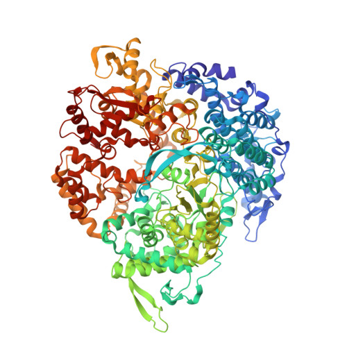
Reovirus Polymerase Lambda3 Localized by Cryo-Electron Microscopy of Virions at a Resolution of 7.6 A

Zhang, X.Walker, S.B.Chipman, P.R.Nibert, M.L.Baker, T.S.

(2003) Nat Struct Biol 10: 1011

  • DOI: https://doi.org/10.1038/nsb1009
  • Primary Citation Related Structures: 
    1UON

  • PubMed Abstract: 

    Reovirus is an icosahedral, double-stranded (ds) RNA virus that uses viral polymerases packaged within the viral core to transcribe its ten distinct plus-strand RNAs. To localize these polymerases, the structure of the reovirion was refined to a resolution of 7.6 A by cryo-electron microscopy (cryo-EM) and three-dimensional (3D) image reconstruction. X-ray crystal models of reovirus proteins, including polymerase lambda 3, were then fitted into the density map. Each copy of lambda 3 was found anchored to the inner surface of the icosahedral core shell, making major contacts with three molecules of shell protein lambda 1 and overlapping, but not centering on, a five-fold axis. The overlap explains why only one copy of lambda 3 is bound per vertex. lambda 3 is furthermore oriented with its transcript exit channel facing a small channel through the lambda 1 shell, suggesting how the nascent RNA is passed into the large external cavity of the pentameric capping enzyme complex formed by protein lambda 2.


  • Organizational Affiliation
    • Department of Biological Sciences, Purdue University, West Lafayette, Indiana 47907, USA.

Macromolecule Content 

  • Total Structure Weight: 148.08 kDa 
  • Atom Count: 10,703 
  • Modeled Residue Count: 1,277 
  • Deposited Residue Count: 1,280 
  • Unique protein chains: 1
  • Unique nucleic acid chains: 2

Macromolecules


Find similar proteins by:|  3D Structure
Entity ID: 1
MoleculeChains  Sequence LengthOrganismDetailsImage
MINOR CORE PROTEIN LAMBDA 31,267Reovirus sp.Mutation(s): 0 
EC: 2.7.7.48
UniProt
Find proteins for P0CK31 (Reovirus type 3 (strain Dearing))
Explore P0CK31 
Go to UniProtKB:  P0CK31
Entity Groups
Sequence Clusters30% Identity50% Identity70% Identity90% Identity95% Identity100% Identity
UniProt GroupP0CK31
Sequence Annotations
Expand
Reference Sequence
Find similar nucleic acids by:  Sequence
Entity ID: 2
MoleculeChains LengthOrganismImage
5'-R(*GP*GP*GP*GP*GP*)-3'5synthetic construct
Sequence Annotations
Expand
Reference Sequence
Find similar nucleic acids by:  Sequence
Entity ID: 3
MoleculeChains LengthOrganismImage
5'-R(*UP*AP*GP*CP*CP*CP*CP*CP*)-3'8synthetic construct
Sequence Annotations
Expand
Reference Sequence

Experimental Data & Validation

Experimental Data

  • Method: ELECTRON MICROSCOPY
  • Resolution: 7.60 Å
  • Aggregation State: PARTICLE 
  • Reconstruction Method: SINGLE PARTICLE 
EM Software:
TaskSoftware PackageVersion
RECONSTRUCTIONPFT

Structure Validation

View Full Validation Report


Entry History 

Deposition Data

Revision History  (Full details and data files)

  • Version 1.0: 2003-11-13
    Type: Initial release
  • Version 1.1: 2011-05-08
    Changes: Version format compliance
  • Version 1.2: 2011-07-13
    Changes: Version format compliance
  • Version 2.0: 2018-10-03
    Changes: Atomic model, Data collection, Derived calculations, Source and taxonomy
  • Version 2.1: 2024-05-08
    Changes: Data collection, Database references, Derived calculations, Refinement description