1UN0 | pdb_00001un0

Crystal Structure of Yeast Karyopherin (Importin) alpha in complex with a Nup2p N-terminal fragment


Experimental Data Snapshot

  • Method: X-RAY DIFFRACTION
  • Resolution: 2.60 Å
  • R-Value Free: 
    0.257 (Depositor) 
  • R-Value Work: 
    0.216 (Depositor) 
  • R-Value Observed: 
    0.218 (Depositor) 

Starting Model: experimental
View more details

wwPDB Validation 3D Report Full Report

Validation slider image for 1UN0

This is version 1.3 of the entry. See complete history

Literature

Structural Basis for Nup2P Function in Cargo Release and Karyopherin Recycling in Nuclear Import

Matsuura, Y.Lange, A.Harreman, M.T.Corbett, A.H.Stewart, M.

(2003) EMBO J 22: 5358

  • DOI: https://doi.org/10.1093/emboj/cdg538
  • Primary Citation Related Structures: 
    1UN0

  • PubMed Abstract: 

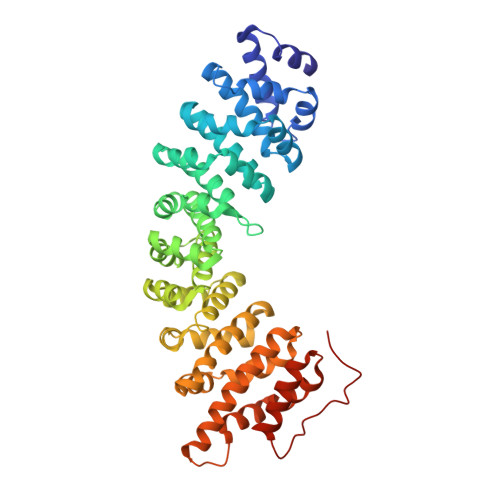
    The yeast nucleoporin Nup2p is associated primarily with the nuclear basket of nuclear pore complexes and is required for efficient importin-alpha:beta-mediated nuclear protein import as well as efficient nuclear export of Kap60p/importin-alpha. Residues 1-51 of Nup2p bind tightly to Kap60p and are required for Nup2p function in vivo. We have determined the 2.6 A resolution crystal structure of a complex between this region of Nup2p and the armadillo repeat domain of Kap60p. Nup2p binds along the inner concave groove of Kap60p, but its interaction interface is different from that employed for nuclear localization signal (NLS) recognition although there is some overlap between them. Nup2p binds Kap60p more strongly than NLSs and accelerates release of NLSs from Kap60p. Nup2p itself is released from Kap60p by Cse1p:RanGTP only in the presence of the importin-beta binding (IBB) domain of Kap60p. These data indicate that Nup2p increases the overall rate of nuclear trafficking by coordinating nuclear import termination and importin recycling as a concerted process.


  • Organizational Affiliation
    • MRC Laboratory of Molecular Biology, Hills Road, Cambridge CB2 2QH, UK.

Macromolecule Content 

  • Total Structure Weight: 109.44 kDa 
  • Atom Count: 7,231 
  • Modeled Residue Count: 911 
  • Deposited Residue Count: 988 
  • Unique protein chains: 2

Macromolecules

Find similar proteins by:|  3D Structure
Entity ID: 1
MoleculeChains  Sequence LengthOrganismDetailsImage
IMPORTIN ALPHA SUBUNIT
A, B
443Saccharomyces cerevisiaeMutation(s): 1 
UniProt
Find proteins for Q02821 (Saccharomyces cerevisiae (strain ATCC 204508 / S288c))
Explore Q02821 
Go to UniProtKB:  Q02821
Entity Groups
Sequence Clusters30% Identity50% Identity70% Identity90% Identity95% Identity100% Identity
UniProt GroupQ02821
Sequence Annotations
Expand
Reference Sequence
Find similar proteins by:|  3D Structure
Entity ID: 2
MoleculeChains  Sequence LengthOrganismDetailsImage
NUCLEOPORIN NUP2
C, D
51Saccharomyces cerevisiaeMutation(s): 0 
UniProt
Find proteins for P32499 (Saccharomyces cerevisiae (strain ATCC 204508 / S288c))
Explore P32499 
Go to UniProtKB:  P32499
Entity Groups
Sequence Clusters30% Identity50% Identity70% Identity90% Identity95% Identity100% Identity
UniProt GroupP32499
Sequence Annotations
Expand
Reference Sequence

Experimental Data & Validation

Experimental Data

  • Method: X-RAY DIFFRACTION
  • Resolution: 2.60 Å
  • R-Value Free:  0.257 (Depositor) 
  • R-Value Work:  0.216 (Depositor) 
  • R-Value Observed: 0.218 (Depositor) 
Space Group: P 21 21 2
Unit Cell:
Length ( Å )Angle ( ˚ )
a = 129.813α = 90
b = 140.076β = 90
c = 63.99γ = 90
Software Package:
Software NamePurpose
REFMACrefinement
MOSFLMdata reduction
SCALAdata scaling
CNSphasing

Structure Validation

View Full Validation Report


Entry History 

Deposition Data

Revision History  (Full details and data files)

  • Version 1.0: 2003-10-16
    Type: Initial release
  • Version 1.1: 2011-05-08
    Changes: Version format compliance
  • Version 1.2: 2011-07-13
    Changes: Version format compliance
  • Version 1.3: 2023-12-13
    Changes: Data collection, Database references, Refinement description