1UMR | pdb_00001umr

Crystal structure of the platelet activator convulxin, a disulfide linked a4b4 cyclic tetramer from the venom of Crotalus durissus terrificus


Experimental Data Snapshot

  • Method: X-RAY DIFFRACTION
  • Resolution: 2.40 Å
  • R-Value Free: 
    0.264 (Depositor), 0.267 (DCC) 
  • R-Value Work: 
    0.187 (Depositor), 0.197 (DCC) 
  • R-Value Observed: 
    0.190 (Depositor) 

Starting Model: experimental
View more details

wwPDB Validation 3D Report Full Report

Validation slider image for 1UMR

This is version 1.3 of the entry. See complete history

Literature

Crystal Structure of the Platelet Activator Convulxin, a Disulfide Linked A4B4 Cyclic Tetramer from the Venom of Crotalus Durissus Terrificus

Murakami, M.T.Zela, S.P.Gava, L.M.Michelan-Duarte, S.Cintra, A.C.O.Arni, R.K.

(2003) Biochem Biophys Res Commun 310: 478

  • DOI: https://doi.org/10.1016/j.bbrc.2003.09.032
  • Primary Citation Related Structures: 
    1UMR

  • PubMed Abstract: 

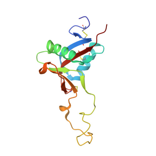
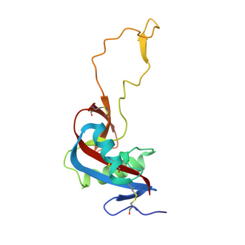
    Convulxin (CVX), a C-type lectin, isolated from the venom of the South American rattlesnake Crotalus durissus terrificus, causes cardiovascular and respiratory disturbances and is a potent platelet activator which binds to platelet glycoprotein GPVI. The structure of CVX has been solved at 2.4A resolution to a crystallographic residual of 18.6% (R(free)=26.4%). CVX is a disulfide linked heterodimer consisting of homologous alpha and beta chains. The heterodimers are additionally linked by disulfide bridges to form cyclic alpha(4)beta(4)heterotetramers. These domains exhibit significant homology to the carbohydrate-binding domains of C-type lectins, to the factor IX-binding protein (IX-bp), and to flavocetin-A (Fl-A) but sequence and structural differences are observed in both the domains in the putative Ca(2+)and carbohydrate binding regions.


  • Organizational Affiliation
    • Department of Physics, IBILCE/UNESP, R. Cristovão Colombo 2265, CEP 15054-000, São Josédo Rio Preto-SP, Brazil.

Macromolecule Content 

  • Total Structure Weight: 61.41 kDa 
  • Atom Count: 4,515 
  • Modeled Residue Count: 520 
  • Deposited Residue Count: 520 
  • Unique protein chains: 2

Macromolecules

Find similar proteins by:|  3D Structure
Entity ID: 1
MoleculeChains  Sequence LengthOrganismDetailsImage
CONVULXIN ALPHA
A, B
135Crotalus durissus terrificusMutation(s): 0 
UniProt
Find proteins for O93426 (Crotalus durissus terrificus)
Explore O93426 
Go to UniProtKB:  O93426
Entity Groups
Sequence Clusters30% Identity50% Identity70% Identity90% Identity95% Identity100% Identity
UniProt GroupO93426
Sequence Annotations
Expand
Reference Sequence
Find similar proteins by:|  3D Structure
Entity ID: 2
MoleculeChains  Sequence LengthOrganismDetailsImage
CONVULXIN BETA
C, D
125Crotalus durissus terrificusMutation(s): 0 
UniProt
Find proteins for O93427 (Crotalus durissus terrificus)
Explore O93427 
Go to UniProtKB:  O93427
Entity Groups
Sequence Clusters30% Identity50% Identity70% Identity90% Identity95% Identity100% Identity
UniProt GroupO93427
Sequence Annotations
Expand
Reference Sequence

Experimental Data & Validation

Experimental Data

  • Method: X-RAY DIFFRACTION
  • Resolution: 2.40 Å
  • R-Value Free:  0.264 (Depositor), 0.267 (DCC) 
  • R-Value Work:  0.187 (Depositor), 0.197 (DCC) 
  • R-Value Observed: 0.190 (Depositor) 
Space Group: I 4
Unit Cell:
Length ( Å )Angle ( ˚ )
a = 131.906α = 90
b = 131.906β = 90
c = 112.849γ = 90
Software Package:
Software NamePurpose
REFMACrefinement
DENZOdata reduction
SCALEPACKdata scaling
AMoREphasing

Structure Validation

View Full Validation Report


Entry History 

Deposition Data

Revision History  (Full details and data files)

  • Version 1.0: 2003-11-21
    Type: Initial release
  • Version 1.1: 2013-11-20
    Changes: Derived calculations, Non-polymer description, Other, Structure summary, Version format compliance
  • Version 1.2: 2023-12-13
    Changes: Data collection, Database references, Other, Refinement description
  • Version 1.3: 2024-10-16
    Changes: Structure summary