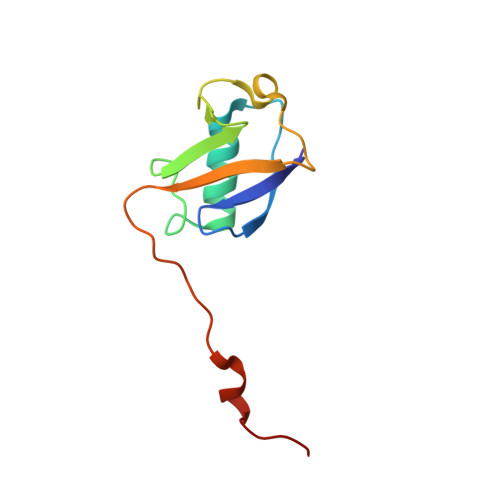
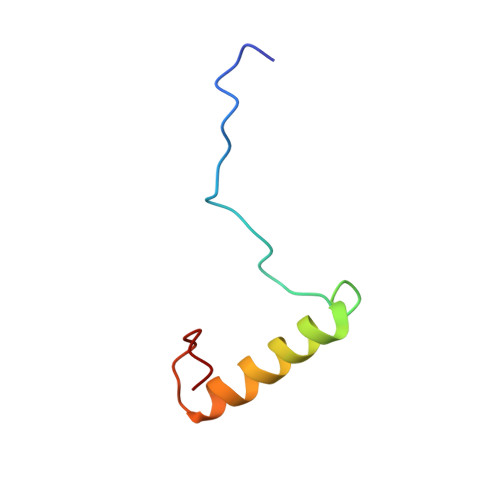
1UEL

Solution structure of ubiquitin-like domain of hHR23B complexed with ubiquitin-interacting motif of proteasome subunit S5a


Experimental Data Snapshot

  • Method: SOLUTION NMR
  • Conformers Calculated: 100 
  • Conformers Submitted: 20 
  • Selection Criteria: The submitted conformer models are the 20 structures with the lowest 

wwPDB Validation   3D Report Full Report


This is version 1.4 of the entry. See complete history


Literature

Structure of the Ubiquitin-interacting Motif of S5a Bound to the Ubiquitin-like Domain of HR23B

Fujiwara, K.Tenno, T.Sugasawa, K.Jee, J.G.Ohki, I.Kojima, C.Tochio, H.Hiroaki, H.Hanaoka, F.Shirakawa, M.

(2004) J Biol Chem 279: 4760-4767

  • DOI: https://doi.org/10.1074/jbc.M309448200
  • Primary Citation of Related Structures:  
    1UEL

  • PubMed Abstract: 

    Ubiquitination, a modification in which single or multiple ubiquitin molecules are attached to a protein, serves signaling functions that control several cellular processes. The ubiquitination signal is recognized by downstream effectors, many of which carry a ubiquitin-interacting motif (UIM). Such interactions can be modulated by regulators carrying a ubiquitin-like (UbL) domain, which binds UIM by mimicking ubiquitination. Of them, HR23B regulates the proteasomal targeting of ubiquitinated substrates, DNA repair factors, and other proteins. Here we report the structure of the UIM of the proteasome subunit S5a bound to the UbL domain of HR23B. The UbL domain presents one hydrophobic and two polar contact sites for interaction with UIM. The residues in these contact sites are well conserved in ubiquitin, but ubiquitin also presents a histidine at the interface. The pH-dependent protonation of this residue interferes with the access of ubiquitin to the UIM and the ubiquitin-associated domain (UBA), and its mutation to a smaller residue increases the affinity of ubiquitin for UIM.


  • Organizational Affiliation

    Graduate School of Integrated Science, Yokohama City University, 1-7-29 Suehiro, Tsurumi, Yokohama, Kanagawa 230-0045, Japan.


Macromolecules
Find similar proteins by:  (by identity cutoff)  |  3D Structure
Entity ID: 1
MoleculeChains Sequence LengthOrganismDetailsImage
UV excision repair protein RAD23 homolog B95Homo sapiensMutation(s): 0 
UniProt & NIH Common Fund Data Resources
Find proteins for P54727 (Homo sapiens)
Explore P54727 
Go to UniProtKB:  P54727
PHAROS:  P54727
GTEx:  ENSG00000119318 
Entity Groups  
Sequence Clusters30% Identity50% Identity70% Identity90% Identity95% Identity100% Identity
UniProt GroupP54727
Sequence Annotations
Expand
  • Reference Sequence
Find similar proteins by:  (by identity cutoff)  |  3D Structure
Entity ID: 2
MoleculeChains Sequence LengthOrganismDetailsImage
26S proteasome non-ATPase regulatory subunit 448Homo sapiensMutation(s): 0 
UniProt & NIH Common Fund Data Resources
Find proteins for P55036 (Homo sapiens)
Explore P55036 
Go to UniProtKB:  P55036
PHAROS:  P55036
GTEx:  ENSG00000159352 
Entity Groups  
Sequence Clusters30% Identity50% Identity70% Identity90% Identity95% Identity100% Identity
UniProt GroupP55036
Sequence Annotations
Expand
  • Reference Sequence
Experimental Data & Validation

Experimental Data

  • Method: SOLUTION NMR
  • Conformers Calculated: 100 
  • Conformers Submitted: 20 
  • Selection Criteria: The submitted conformer models are the 20 structures with the lowest 

Structure Validation

View Full Validation Report


Entry History 

Revision History  (Full details and data files)

  • Version 1.0: 2004-02-10
    Type: Initial release
  • Version 1.1: 2008-04-27
    Changes: Version format compliance
  • Version 1.2: 2011-07-13
    Changes: Version format compliance
  • Version 1.3: 2022-03-02
    Changes: Database references, Derived calculations
  • Version 1.4: 2023-12-27
    Changes: Data collection