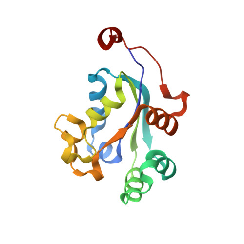
1UCN

X-ray structure of human nucleoside diphosphate kinase A complexed with ADP at 2 A resolution


Experimental Data Snapshot

  • Method: X-RAY DIFFRACTION
  • Resolution: 2.00 Å
  • R-Value Free: 0.282 
  • R-Value Work: 0.237 

wwPDB Validation   3D Report Full Report


Ligand Structure Quality Assessment 


This is version 1.4 of the entry. See complete history


Literature

Nucleotide Binding to Nucleoside Diphosphate Kinases: X-ray Structure of Human NDPK-A in Complex with ADP and Comparison to Protein Kinases

Chen, Y.Gallois-Montbrun, S.Schneider, B.Veron, M.Morera, S.Deville-Bonne, D.Janin, J.

(2003) J Mol Biol 332: 915-926

  • DOI: https://doi.org/10.1016/j.jmb.2003.07.004
  • Primary Citation of Related Structures:  
    1UCN

  • PubMed Abstract: 

    NDPK-A, product of the nm23-H1 gene, is one of the two major isoforms of human nucleoside diphosphate kinase. We analyzed the binding of its nucleotide substrates by fluorometric methods. The binding of nucleoside triphosphate (NTP) substrates was detected by following changes of the intrinsic fluorescence of the H118G/F60W variant, a mutant protein engineered for that purpose. Nucleoside diphosphate (NDP) substrate binding was measured by competition with a fluorescent derivative of ADP, following the fluorescence anisotropy of the derivative. We also determined an X-ray structure at 2.0A resolution of the variant NDPK-A in complex with ADP, Ca(2+) and inorganic phosphate, products of ATP hydrolysis. We compared the conformation of the bound nucleotide seen in this complex and the interactions it makes with the protein, with those of the nucleotide substrates, substrate analogues or inhibitors present in other NDP kinase structures. We also compared NDP kinase-bound nucleotides to ATP bound to protein kinases, and showed that the nucleoside monophosphate moieties have nearly identical conformations in spite of the very different protein environments. However, the beta and gamma-phosphate groups are differently positioned and oriented in the two types of kinases, and they bind metal ions with opposite chiralities. Thus, it should be possible to design nucleotide analogues that are good substrates of one type of kinase, and poor substrates or inhibitors of the other kind.


  • Organizational Affiliation

    Laboratoire d'Enzymologie et Biochimie Structurales, CNRS UPR 9063, 91198 Gif-sur Yvette, France.


Macromolecules
Find similar proteins by:  (by identity cutoff)  |  3D Structure
Entity ID: 1
MoleculeChains Sequence LengthOrganismDetailsImage
nucleoside diphosphate kinase A
A, B, C
152Homo sapiensMutation(s): 2 
EC: 2.7.4.6
UniProt & NIH Common Fund Data Resources
Find proteins for P15531 (Homo sapiens)
Explore P15531 
Go to UniProtKB:  P15531
PHAROS:  P15531
GTEx:  ENSG00000239672 
Entity Groups  
Sequence Clusters30% Identity50% Identity70% Identity90% Identity95% Identity100% Identity
UniProt GroupP15531
Sequence Annotations
Expand
  • Reference Sequence
Small Molecules
Ligands 4 Unique
IDChains Name / Formula / InChI Key2D Diagram3D Interactions
ADP
Query on ADP

Download Ideal Coordinates CCD File 
F [auth A],
J [auth B],
O [auth C]
ADENOSINE-5'-DIPHOSPHATE
C10 H15 N5 O10 P2
XTWYTFMLZFPYCI-KQYNXXCUSA-N
TRS
Query on TRS

Download Ideal Coordinates CCD File 
K [auth B]2-AMINO-2-HYDROXYMETHYL-PROPANE-1,3-DIOL
C4 H12 N O3
LENZDBCJOHFCAS-UHFFFAOYSA-O
PO4
Query on PO4

Download Ideal Coordinates CCD File 
D [auth A],
G [auth B],
L [auth C]
PHOSPHATE ION
O4 P
NBIIXXVUZAFLBC-UHFFFAOYSA-K
CA
Query on CA

Download Ideal Coordinates CCD File 
E [auth A],
H [auth B],
I [auth B],
M [auth C],
N [auth C]
CALCIUM ION
Ca
BHPQYMZQTOCNFJ-UHFFFAOYSA-N
Binding Affinity Annotations 
IDSourceBinding Affinity
ADP PDBBind:  1UCN Kd: 6000 (nM) from 1 assay(s)
Experimental Data & Validation

Experimental Data

  • Method: X-RAY DIFFRACTION
  • Resolution: 2.00 Å
  • R-Value Free: 0.282 
  • R-Value Work: 0.237 
  • Space Group: P 41 21 2
Unit Cell:
Length ( Å )Angle ( ˚ )
a = 116.075α = 90
b = 116.075β = 90
c = 91.292γ = 90
Software Package:
Software NamePurpose
DENZOdata reduction
SCALEPACKdata scaling
CNSrefinement
CNSphasing

Structure Validation

View Full Validation Report


Ligand Structure Quality Assessment 


Entry History 

Deposition Data

Revision History  (Full details and data files)

  • Version 1.0: 2003-09-30
    Type: Initial release
  • Version 1.1: 2008-04-27
    Changes: Version format compliance
  • Version 1.2: 2011-07-13
    Changes: Derived calculations, Version format compliance
  • Version 1.3: 2021-11-10
    Changes: Database references, Derived calculations
  • Version 1.4: 2023-12-27
    Changes: Data collection