1U04 | pdb_00001u04

Crystal structure of full length Argonaute from Pyrococcus furiosus


Experimental Data Snapshot

  • Method: X-RAY DIFFRACTION
  • Resolution: 2.25 Å
  • R-Value Free: 
    0.258 (Depositor), 0.261 (DCC) 
  • R-Value Work: 
    0.227 (Depositor), 0.231 (DCC) 
  • R-Value Observed: 
    0.227 (Depositor) 

wwPDB Validation 3D Report Full Report

Validation slider image for 1U04

This is version 1.5 of the entry. See complete history

Literature

Crystal structure of Argonaute and its implications for RISC slicer activity.

Song, J.J.Smith, S.K.Hannon, G.J.Joshua-Tor, L.

(2004) Science 305: 1434-1437

  • DOI: https://doi.org/10.1126/science.1102514
  • Primary Citation Related Structures: 
    1U04

  • PubMed Abstract: 

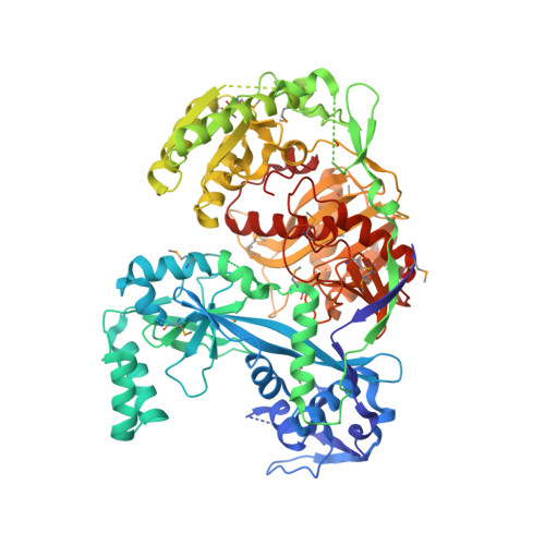
    Argonaute proteins and small interfering RNAs (siRNAs) are the known signature components of the RNA interference effector complex RNA-induced silencing complex (RISC). However, the identity of "Slicer," the enzyme that cleaves the messenger RNA (mRNA) as directed by the siRNA, has not been resolved. Here, we report the crystal structure of the Argonaute protein from Pyrococcus furiosus at 2.25 angstrom resolution. The structure reveals a crescent-shaped base made up of the amino-terminal, middle, and PIWI domains. The Piwi Argonaute Zwille (PAZ) domain is held above the base by a "stalk"-like region. The PIWI domain (named for the protein piwi) is similar to ribonuclease H, with a conserved active site aspartate-aspartate-glutamate motif, strongly implicating Argonaute as "Slicer." The architecture of the molecule and the placement of the PAZ and PIWI domains define a groove for substrate binding and suggest a mechanism for siRNA-guided mRNA cleavage.


  • Organizational Affiliation
    • Watson School of Biological Sciences, 1 Bungtown Road, Cold Spring Harbor, NY 11724, USA.

Macromolecule Content 

  • Total Structure Weight: 91.44 kDa 
  • Atom Count: 6,112 
  • Modeled Residue Count: 713 
  • Deposited Residue Count: 771 
  • Unique protein chains: 1

Macromolecules

Find similar proteins by:|  3D Structure
Entity ID: 1
MoleculeChains  Sequence LengthOrganismDetailsImage
hypothetical protein PF0537771Pyrococcus furiosus DSM 3638Mutation(s): 1 
EC: 3.1.24
UniProt
Find proteins for Q8U3D2 (Pyrococcus furiosus (strain ATCC 43587 / DSM 3638 / JCM 8422 / Vc1))
Explore Q8U3D2 
Go to UniProtKB:  Q8U3D2
Entity Groups
Sequence Clusters30% Identity50% Identity70% Identity90% Identity95% Identity100% Identity
UniProt GroupQ8U3D2
Sequence Annotations
Expand
Reference Sequence

Small Molecules

Modified Residues  1 Unique
IDChains TypeFormula2D DiagramParent
MSE
Query on MSE
A
L-PEPTIDE LINKINGC5 H11 N O2 SeMET

Experimental Data & Validation

Experimental Data

  • Method: X-RAY DIFFRACTION
  • Resolution: 2.25 Å
  • R-Value Free:  0.258 (Depositor), 0.261 (DCC) 
  • R-Value Work:  0.227 (Depositor), 0.231 (DCC) 
  • R-Value Observed: 0.227 (Depositor) 
Space Group: P 1 21 1
Unit Cell:
Length ( Å )Angle ( ˚ )
a = 69.726α = 90
b = 104.188β = 102.83
c = 74.015γ = 90
Software Package:
Software NamePurpose
CNSrefinement
HKL-2000data reduction
SCALEPACKdata scaling
SHARPphasing
SnBphasing

Structure Validation

View Full Validation Report


Entry History 

Deposition Data

Revision History  (Full details and data files)

  • Version 1.0: 2004-08-17
    Type: Initial release
  • Version 1.1: 2008-04-30
    Changes: Version format compliance
  • Version 1.2: 2011-07-13
    Changes: Source and taxonomy, Version format compliance
  • Version 1.3: 2021-10-27
    Changes: Database references, Derived calculations
  • Version 1.4: 2024-10-30
    Changes: Data collection, Structure summary
  • Version 1.5: 2026-02-11
    Changes: Data collection, Database references