1TZY

Crystal Structure of the Core-Histone Octamer to 1.90 Angstrom Resolution


Experimental Data Snapshot

  • Method: X-RAY DIFFRACTION
  • Resolution: 1.90 Å
  • R-Value Free: 0.222 
  • R-Value Work: 0.187 
  • R-Value Observed: 0.188 

wwPDB Validation   3D Report Full Report


This is version 1.3 of the entry. See complete history


Literature

High-resolution structure of the native histone octamer.

Wood, C.M.Nicholson, J.M.Lambert, S.J.Chantalat, L.Reynolds, C.D.Baldwin, J.P.

(2005) Acta Crystallogr Sect F Struct Biol Cryst Commun 61: 541-545

  • DOI: https://doi.org/10.1107/S1744309105013813
  • Primary Citation of Related Structures:  
    1TZY

  • PubMed Abstract: 

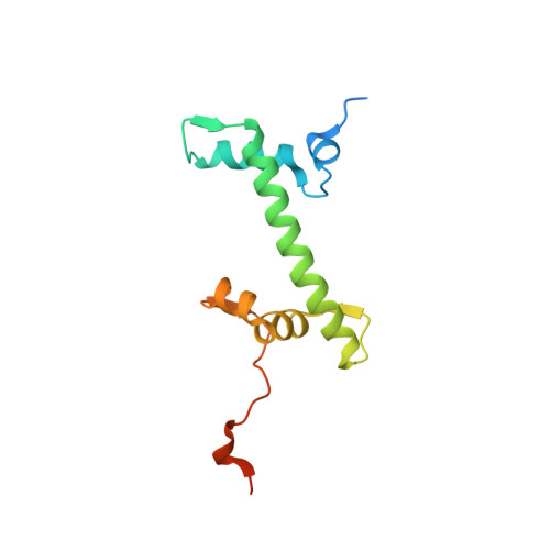
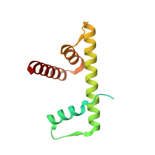
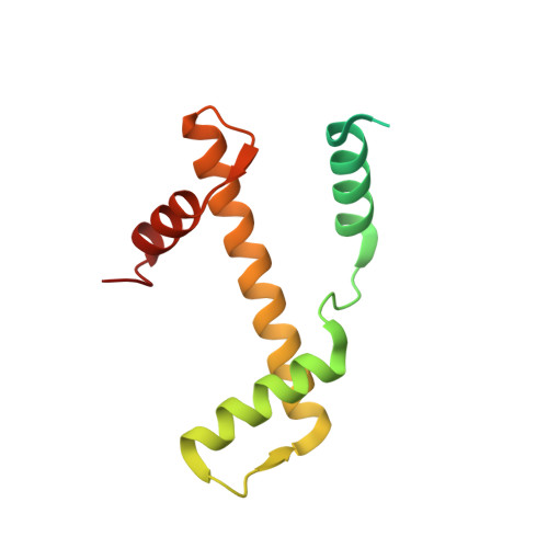
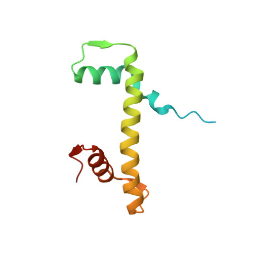
    Crystals of native histone octamers (H2A-H2B)-(H4-H3)-(H3'-H4')-(H2B'-H2A') from chick erythrocytes in 2 M KCl, 1.35 M potassium phosphate pH 6.9 diffract X-rays to 1.90 A resolution, yielding a structure with an R(work) value of 18.7% and an Rfree of 22.2%. The crystal space group is P6(5), the asymmetric unit of which contains one complete octamer. This high-resolution model of the histone-core octamer allows further insight into intermolecular interactions, including water molecules, that dock the histone dimers to the tetramer in the nucleosome-core particle and have relevance to nucleosome remodelling. The three key areas analysed are the H2A'-H3-H4 molecular cluster (also H2A-H3'-H4'), the H4-H2B' interaction (also H4'-H2B) and the H2A'-H4 beta-sheet interaction (also H2A-H4'). The latter of these three regions is important to nucleosome remodelling by RNA polymerase II, as it is shown to be a likely core-histone binding site, and its disruption creates an instability in the nucleosome-core particle. A majority of the water molecules in the high-resolution octamer have positions that correlate to similar positions in the high-resolution nucleosome-core particle structure, suggesting that the high-resolution octamer model can be used for comparative studies with the high-resolution nucleosome-core particle.


  • Organizational Affiliation

    School of Biomolecular Sciences, Liverpool John Moores University, Liverpool L3 3AF, England. c.m.wood@livjm.ac.uk


Macromolecules
Find similar proteins by:  (by identity cutoff)  |  3D Structure
Entity ID: 1
MoleculeChains Sequence LengthOrganismDetailsImage
Histone H2A-IV
A, E
129Gallus gallusMutation(s): 0 
UniProt
Find proteins for P02263 (Gallus gallus)
Explore P02263 
Go to UniProtKB:  P02263
Entity Groups  
Sequence Clusters30% Identity50% Identity70% Identity90% Identity95% Identity100% Identity
UniProt GroupP02263
Sequence Annotations
Expand
  • Reference Sequence
Find similar proteins by:  (by identity cutoff)  |  3D Structure
Entity ID: 2
MoleculeChains Sequence LengthOrganismDetailsImage
Histone H2B
B, F
126Gallus gallusMutation(s): 0 
UniProt
Find proteins for P0C1H5 (Gallus gallus)
Explore P0C1H5 
Go to UniProtKB:  P0C1H5
Entity Groups  
Sequence Clusters30% Identity50% Identity70% Identity90% Identity95% Identity100% Identity
UniProt GroupP0C1H5
Sequence Annotations
Expand
  • Reference Sequence
Find similar proteins by:  (by identity cutoff)  |  3D Structure
Entity ID: 3
MoleculeChains Sequence LengthOrganismDetailsImage
HISTONE H3
C, G
136Gallus gallusMutation(s): 0 
UniProt
Find proteins for P84229 (Gallus gallus)
Explore P84229 
Go to UniProtKB:  P84229
Entity Groups  
Sequence Clusters30% Identity50% Identity70% Identity90% Identity95% Identity100% Identity
UniProt GroupP84229
Sequence Annotations
Expand
  • Reference Sequence
Find similar proteins by:  (by identity cutoff)  |  3D Structure
Entity ID: 4
MoleculeChains Sequence LengthOrganismDetailsImage
HISTONE H4-VI
D, H
103Gallus gallusMutation(s): 0 
UniProt
Find proteins for P62801 (Gallus gallus)
Explore P62801 
Go to UniProtKB:  P62801
Entity Groups  
Sequence Clusters30% Identity50% Identity70% Identity90% Identity95% Identity100% Identity
UniProt GroupP62801
Sequence Annotations
Expand
  • Reference Sequence
Small Molecules
Ligands 2 Unique
IDChains Name / Formula / InChI Key2D Diagram3D Interactions
PO4
Query on PO4

Download Ideal Coordinates CCD File 
I [auth A],
N [auth B],
Q [auth C],
X [auth E],
Y [auth E]
PHOSPHATE ION
O4 P
NBIIXXVUZAFLBC-UHFFFAOYSA-K
CL
Query on CL

Download Ideal Coordinates CCD File 
AA [auth E]
BA [auth F]
CA [auth F]
DA [auth G]
EA [auth G]
AA [auth E],
BA [auth F],
CA [auth F],
DA [auth G],
EA [auth G],
FA [auth G],
GA [auth G],
HA [auth H],
IA [auth H],
J [auth A],
JA [auth H],
K [auth A],
L [auth A],
M [auth A],
O [auth B],
P [auth B],
R [auth C],
S [auth C],
T [auth C],
U [auth D],
V [auth D],
W [auth D],
Z [auth E]
CHLORIDE ION
Cl
VEXZGXHMUGYJMC-UHFFFAOYSA-M
Experimental Data & Validation

Experimental Data

  • Method: X-RAY DIFFRACTION
  • Resolution: 1.90 Å
  • R-Value Free: 0.222 
  • R-Value Work: 0.187 
  • R-Value Observed: 0.188 
  • Space Group: P 65
Unit Cell:
Length ( Å )Angle ( ˚ )
a = 158.351α = 90
b = 158.351β = 90
c = 103.576γ = 120
Software Package:
Software NamePurpose
REFMACrefinement
PXGENdata reduction
SCALEPACKdata scaling
AMoREphasing

Structure Validation

View Full Validation Report


Entry History 

Deposition Data

Revision History  (Full details and data files)

  • Version 1.0: 2004-08-03
    Type: Initial release
  • Version 1.1: 2008-04-30
    Changes: Version format compliance
  • Version 1.2: 2011-07-13
    Changes: Version format compliance
  • Version 1.3: 2024-03-13
    Changes: Data collection, Database references, Derived calculations