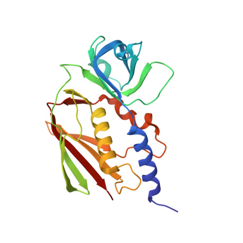
Crystallographic and mutational data show that the streptococcal pyrogenic exotoxin j can use a common binding surface for T-cell receptor binding and dimerization
Baker, H.M., Proft, T., Webb, P.D., Arcus, V.L., Fraser, J.D., Baker, E.N.(2004) J Biological Chem 279: 38571-38576
- PubMed: 15247241
- DOI: https://doi.org/10.1074/jbc.M406695200
- Primary Citation of Related Structures:
1TY0, 1TY2 - PubMed Abstract:
The protein toxins known as superantigens (SAgs), which are expressed primarily by the pathogenic bacteria Staphylococcus aureus and Streptococcus pyogenes, are highly potent immunotoxins with the ability to cause serious human disease. These SAgs share a conserved fold but quite varied activities. In addition to their common role of cross-linking T-cell receptors (TCRs) and major histocompatibility complex class II (MHC-II) molecules, some SAgs can cross-link MHC-II, using diverse mechanisms. The crystal structure of the streptococcal superantigen streptococcal pyrogenic exotoxin J (SPE-J) has been solved at 1.75 A resolution (R = 0.209, R(free) = 0.240), both with and without bound Zn(2+). The structure displays the canonical two-domain SAg fold and a zinc-binding site that is shared by a subset of other SAgs. Most importantly, in concentrated solution and in the crystal, SPE-J forms dimers. These dimers, which are present in two different crystal environments, form via the same face that is used for TCR binding in other SAgs. Site-directed mutagenesis shows that this face is also used for TCR binding SPE-J. We infer that SPE-J cross-links TCR and MHC-II as a monomer but that dimers may form on the antigen-presenting cell surface, cross-linking MHC-II and eliciting intracellular signaling.
- School of Biological Sciences, University of Auckland, Private Bag 92019, Auckland, New Zealand.
Organizational Affiliation:


