X-ray and thermodynamic studies of staphylococcal nuclease variants I92E and I92K: insights into polarity of the protein interior
Nguyen, D.M., Reynald, R.L., Gittis, A.G., Lattman, E.E.(2004) J Mol Biol 341: 565-574
- PubMed: 15276844 Search on PubMed
- DOI: https://doi.org/10.1016/j.jmb.2004.05.066
- Primary Citation Related Structures:
1TQO, 1TR5, 1TT2 - PubMed Abstract:
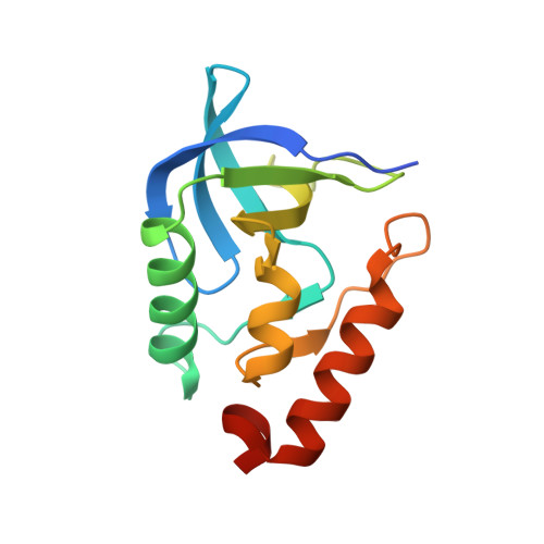
We have used crystallography and thermodynamic analysis to study nuclease variants I92E and I92K, in which an ionizable side-chain is placed in the hydrophobic core of nuclease. We find that the energetic cost of burying ionizable groups is rather modest. The X-ray determinations show water molecules solvating the buried glutamic acid under cryo conditions, but not at room temperature. The lysine side-chain does not appear solvated in either case. Guanidine hydrochloride (GnHCl) denaturation of I92E and I92K, done as a function of pH and monitored by tryptophan fluorescence, showed that I92E and I92K are folded in the pH range pH 3.5-9.0 and pH 5.5-9.5, respectively. The stability of the parental protein is independent of pH over a broad range. In contrast, the stabilities of I92E and I92K exhibit a pH dependence, which is quantitatively explained by thermodynamic analysis: the PK(a) value of the buried K92 is 5.6, while that of the buried E92 is 8.65. The free energy difference between burying the uncharged and charged forms of the groups is modest, about 6 kcal/mol. We also found that epsilon(app) for I92K and I92E is in the range approximately 10-12, instead of 2-4 commonly used to represent the protein interior. Side-chains 92E and 92K were uncharged under the conditions of the X-ray experiment. Both are buried completely inside the well-defined hydrophobic core of the variant proteins without forming salt-bridges or hydrogen bonds to other functional groups of the proteins. Under cryo conditions 92E shows a chain of four water molecules, which hydrate one oxygen atom of the carboxyl group of the glutamic acid. Two other water molecules, which are present in the wild-type at all temperatures, are also connected to the water ring observed inside the hydrophobic core. The ready burial of water with an uncharged E92 raises the possibility that solvent excursions into the interior also take place in the wild-type protein, but in a random, dynamic way not detectable by crystallography. Such transient excursions could increase the average polarity, and thus epsilon(app), of the protein interior.
- Department of Biophysics, Johns Hopkins University, 3400 North Charles Street, Baltimore, MD 21218, USA.
Organizational Affiliation: