1TT0

Crystal Structure of Pyranose 2-Oxidase


Experimental Data Snapshot

  • Method: X-RAY DIFFRACTION
  • Resolution: 1.80 Å
  • R-Value Free: 0.171 
  • R-Value Work: 0.134 
  • R-Value Observed: 0.134 

wwPDB Validation   3D Report Full Report


Ligand Structure Quality Assessment 


This is version 1.4 of the entry. See complete history


Literature

Crystal structure of the 270 kDa homotetrameric lignin-degrading enzyme pyranose 2-oxidase

Hallberg, B.M.Leitner, C.Haltrich, D.Divne, C.

(2004) J Mol Biol 341: 781-796

  • DOI: https://doi.org/10.1016/j.jmb.2004.06.033
  • Primary Citation of Related Structures:  
    1TT0

  • PubMed Abstract: 

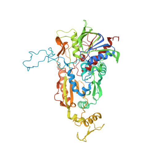
    Pyranose 2-oxidase (P2Ox) is a 270 kDa homotetramer localized preferentially in the hyphal periplasmic space of lignocellulolytic fungi and has a proposed role in lignocellulose degradation to produce the essential co-substrate, hydrogen peroxide, for lignin peroxidases. P2Ox oxidizes D-glucose and other aldopyranoses regioselectively at C2 to the corresponding 2-keto sugars; however, for some substrates, the enzyme also displays specificity for oxidation at C3. The crystal structure of P2Ox from Trametes multicolor has been determined using single anomalous dispersion with mercury as anomalous scatterer. The model was refined at 1.8A resolution to R and Rfree values of 0.134 and 0.171, respectively. The overall fold of the P2Ox subunit resembles that of members of the glucose-methanol-choline family of long-chain oxidoreductases, featuring a flavin-binding Rossmann domain of class alpha/beta and a substrate-binding subdomain with a six-stranded central beta sheet and three alpha helices. The homotetramer buries a large internal cavity of roughly 15,000 A3, from which the four active sites are accessible. Four solvent channels lead from the surface into the cavity through which substrate must enter before accessing the active site. The present structure shows an acetate molecule bound in the active site with the carboxylate group positioned immediately below the flavin N5 atom, and with one carboxylate oxygen atom interacting with the catalytic residues His548 and Asn593. The entrance to the active site is blocked by a loop (residues 452 to 461) with excellent electron density but elevated temperature factors. We predict that this loop is dynamic and opens to allow substrate entry and exit. In silico docking of D-glucose in the P2Ox active site shows that with the active-site loop in the closed conformation, monosaccharides cannot be accommodated; however, after removing the loop from the model, a tentative set of protein-substrate interactions for beta-D-glucose have been outlined.


  • Organizational Affiliation

    Department of Biotechnology, KTH, Albanova University Center, SE-106 91 Stockholm, Sweden.


Macromolecules
Find similar proteins by:  (by identity cutoff)  |  3D Structure
Entity ID: 1
MoleculeChains Sequence LengthOrganismDetailsImage
pyranose oxidase
A, B, C, D
623Trametes ochraceaMutation(s): 0 
EC: 1.1.3.10
UniProt
Find proteins for Q7ZA32 (Trametes ochracea)
Explore Q7ZA32 
Go to UniProtKB:  Q7ZA32
Entity Groups  
Sequence Clusters30% Identity50% Identity70% Identity90% Identity95% Identity100% Identity
UniProt GroupQ7ZA32
Sequence Annotations
Expand
  • Reference Sequence
Small Molecules
Ligands 3 Unique
IDChains Name / Formula / InChI Key2D Diagram3D Interactions
FAD
Query on FAD

Download Ideal Coordinates CCD File 
AA [auth D],
F [auth A],
M [auth B],
U [auth C]
FLAVIN-ADENINE DINUCLEOTIDE
C27 H33 N9 O15 P2
VWWQXMAJTJZDQX-UYBVJOGSSA-N
12P
Query on 12P

Download Ideal Coordinates CCD File 
BA [auth D]
CA [auth D]
DA [auth D]
EA [auth D]
FA [auth D]
BA [auth D],
CA [auth D],
DA [auth D],
EA [auth D],
FA [auth D],
G [auth A],
H [auth A],
I [auth A],
J [auth A],
K [auth A],
N [auth B],
O [auth B],
P [auth B],
Q [auth B],
R [auth B],
S [auth B],
V [auth C],
W [auth C],
X [auth C],
Y [auth C]
DODECAETHYLENE GLYCOL
C24 H50 O13
WRZXKWFJEFFURH-UHFFFAOYSA-N
ACT
Query on ACT

Download Ideal Coordinates CCD File 
E [auth A],
L [auth B],
T [auth C],
Z [auth D]
ACETATE ION
C2 H3 O2
QTBSBXVTEAMEQO-UHFFFAOYSA-M
Experimental Data & Validation

Experimental Data

  • Method: X-RAY DIFFRACTION
  • Resolution: 1.80 Å
  • R-Value Free: 0.171 
  • R-Value Work: 0.134 
  • R-Value Observed: 0.134 
  • Space Group: P 1 21 1
Unit Cell:
Length ( Å )Angle ( ˚ )
a = 99.92α = 90
b = 101.69β = 90.95
c = 135.63γ = 90
Software Package:
Software NamePurpose
REFMACrefinement
XDSdata scaling
SOLVEphasing

Structure Validation

View Full Validation Report


Ligand Structure Quality Assessment 


Entry History 

Deposition Data

Revision History  (Full details and data files)

  • Version 1.0: 2005-06-21
    Type: Initial release
  • Version 1.1: 2008-04-30
    Changes: Version format compliance
  • Version 1.2: 2011-07-13
    Changes: Advisory, Version format compliance
  • Version 1.3: 2014-11-12
    Changes: Structure summary
  • Version 1.4: 2024-02-14
    Changes: Advisory, Data collection, Database references, Derived calculations