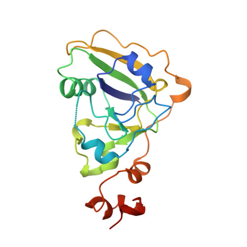
1TII

ESCHERICHIA COLI HEAT LABILE ENTEROTOXIN TYPE IIB


Experimental Data Snapshot

  • Method: X-RAY DIFFRACTION
  • Resolution: 2.25 Å
  • R-Value Free: 0.266 
  • R-Value Work: 0.191 
  • R-Value Observed: 0.191 

wwPDB Validation   3D Report Full Report


This is version 1.2 of the entry. See complete history


Literature

Crystal structure of a new heat-labile enterotoxin, LT-IIb.

van den Akker, F.Sarfaty, S.Twiddy, E.M.Connell, T.D.Holmes, R.K.Hol, W.G.

(1996) Structure 4: 665-678

  • DOI: https://doi.org/10.1016/s0969-2126(96)00073-1
  • Primary Citation of Related Structures:  
    1TII

  • PubMed Abstract: 

    Cholera toxin from Vibrio cholerae and the type I heat-labile enterotoxins (LT-Is) from Escherichia coli are oligomeric proteins with AB5 structures. The type II heat-labile enterotoxins (LT-IIs) from E. coli are structurally similar to, but antigenically distinct from, the type I enterotoxins. The A subunits of type I and type II enterotoxins are homologous and activate adenylate cyclase by ADP-ribosylation of a G protein subunit, G8 alpha. However, the B subunits of type I and type II enterotoxins differ dramatically in amino acid sequence and ganglioside-binding specificity. The structure of LT-IIb was determined both as a prototype for other LT-IIs and to provide additional insights into structure/function relationships among members of the heat-labile enterotoxin family and the superfamily of ADP-ribosylating protein toxins. The 2.25 A crystal structure of the LT-IIb holotoxin has been determined. The structure reveals striking similarities with LT-I in both the catalytic A subunit and the ganglioside-binding B subunits. The latter form a pentamer which has a central pore with a diameter of 10-18 A. Despite their similarities, the relative orientation between the A polypeptide and the B pentamer differs by 24 degrees in LT-I and LT-IIb. A common hydrophobic ring was observed at the A-B5 interface which may be important in the cholera toxin family for assembly of the AB5 heterohexamer. A cluster of arginine residues at the surface of the A subunit of LT-I and cholera toxin, possibly involved in assembly, is also present in LT-IIb. The ganglioside receptor binding sites are localized, as suggested by mutagenesis, and are in a position roughly similar to the sites where LT-I binds its receptor. The structure of LT-IIb provides insight into the sequence diversity and structural similarity of the AB5 toxin family. New knowledge has been gained regarding the assembly of AB5 toxins and their active-site architecture.


  • Organizational Affiliation

    Department of Biological Structure and Biochemistry, University of Washington, Seattle 98195, USA.


Macromolecules
Find similar proteins by:  (by identity cutoff)  |  3D Structure
Entity ID: 1
MoleculeChains Sequence LengthOrganismDetailsImage
HEAT LABILE ENTEROTOXIN TYPE IIBA [auth D],
B [auth E],
C [auth F],
D [auth G],
E [auth H]
99Escherichia coliMutation(s): 0 
UniProt
Find proteins for P43529 (Escherichia coli)
Explore P43529 
Go to UniProtKB:  P43529
Entity Groups  
Sequence Clusters30% Identity50% Identity70% Identity90% Identity95% Identity100% Identity
UniProt GroupP43529
Sequence Annotations
Expand
  • Reference Sequence
Find similar proteins by:  (by identity cutoff)  |  3D Structure
Entity ID: 2
MoleculeChains Sequence LengthOrganismDetailsImage
HEAT LABILE ENTEROTOXIN TYPE IIBF [auth A]190Escherichia coliMutation(s): 0 
UniProt
Find proteins for P43528 (Escherichia coli)
Explore P43528 
Go to UniProtKB:  P43528
Entity Groups  
Sequence Clusters30% Identity50% Identity70% Identity90% Identity95% Identity100% Identity
UniProt GroupP43528
Sequence Annotations
Expand
  • Reference Sequence
Find similar proteins by:  (by identity cutoff)  |  3D Structure
Entity ID: 3
MoleculeChains Sequence LengthOrganismDetailsImage
HEAT LABILE ENTEROTOXIN TYPE IIBG [auth C]53Escherichia coliMutation(s): 0 
UniProt
Find proteins for P43528 (Escherichia coli)
Explore P43528 
Go to UniProtKB:  P43528
Entity Groups  
Sequence Clusters30% Identity50% Identity70% Identity90% Identity95% Identity100% Identity
UniProt GroupP43528
Sequence Annotations
Expand
  • Reference Sequence
Experimental Data & Validation

Experimental Data

  • Method: X-RAY DIFFRACTION
  • Resolution: 2.25 Å
  • R-Value Free: 0.266 
  • R-Value Work: 0.191 
  • R-Value Observed: 0.191 
  • Space Group: P 31 2 1
Unit Cell:
Length ( Å )Angle ( ˚ )
a = 105.7α = 90
b = 105.7β = 90
c = 171.6γ = 120
Software Package:
Software NamePurpose
DENZOdata reduction
SCALEPACKdata scaling
X-PLORmodel building
X-PLORrefinement
X-PLORphasing

Structure Validation

View Full Validation Report


Entry History 

Deposition Data

Revision History  (Full details and data files)

  • Version 1.0: 1996-08-17
    Type: Initial release
  • Version 1.1: 2008-03-24
    Changes: Version format compliance
  • Version 1.2: 2011-07-13
    Changes: Version format compliance